Scolaris Content Display Scolaris Content Display

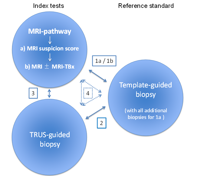
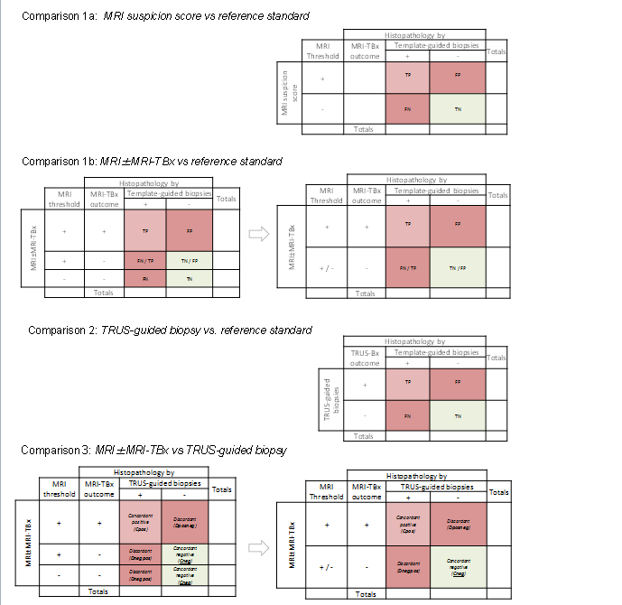
MRI pathway and TRUS‐guided biopsy for detecting clinically significant prostate cancer

Esta versión no es la más reciente

The MRI pathway comprises two components: MRI (suspicion score) and MRI‐targeted biopsy in the presence of suspicious lesions for clinically significant prostate cancer:a) MRI suspicion score: refers to only the first component of the MRI pathway. b) MRI±MRI‐TBx: refers to both components of the MRI pathway. Review design Meta‐analysis of studies reporting on:The MRI pathway and the reference standard: ·Comparison 1a: MRI suspicion score vs reference standard and all additional biopsies (e.g. MRI‐targeted biopsies). Comparison 1b: MRI±MRI‐TBx vs reference standard. TRUS‐guided biopsy and reference standard: ·Comparison 2 MRI pathway B) MRI±MRI‐TBx and TRUS‐guide biopsy: Comparison 3 A combination of all tests: Comparison 4: comparing both index tests, either directly with the reference standard or indirectly as a randomised trial of test accuracy.
Figuras y tablas -
Figure 1

The MRI pathway comprises two components: MRI (suspicion score) and MRI‐targeted biopsy in the presence of suspicious lesions for clinically significant prostate cancer:

  • a) MRI suspicion score: refers to only the first component of the MRI pathway.

  • b) MRI±MRI‐TBx: refers to both components of the MRI pathway.

Review design

Meta‐analysis of studies reporting on:

The MRI pathway and the reference standard: ·

  • Comparison 1a: MRI suspicion score vs reference standard and all additional biopsies (e.g. MRI‐targeted biopsies).

  • Comparison 1b: MRI±MRI‐TBx vs reference standard.

TRUS‐guided biopsy and reference standard: ·

  • Comparison 2

MRI pathway B) MRI±MRI‐TBx and TRUS‐guide biopsy:

  • Comparison 3

A combination of all tests:

  • Comparison 4: comparing both index tests, either directly with the reference standard or indirectly as a randomised trial of test accuracy.

Two‐by‐two contingency tables for target condition.Comparison 1a: MRI suspicion score vs reference standard (if MRI‐targeted biopsies are performed these will serve as additional reference biopsies).Comparison 1b: MRI±MRI‐TBx vs reference standard.Comparison 2: TRUS‐guided biopsy vs reference standard.Comparison 3: MRI±MRI‐TBx vs TRUS‐guided biopsy.For comparison 1b and comparison 3, a negative test can result in two ways:a negative MRI (thus no MRI‐targeted biopsies are taken). a positive MRI but negative MRI‐targeted biopsy result. Both negative outcomes should be merged, creating a two‐by‐two from a three‐by‐two contingency table (first and second column).In comparison 3 the TRUS‐guided biopsy is not a proper reference standard. Therefore we will focus on the number of cancers identified and the concordance and disconcordance between both index tests.TP = true positive; FP = false positive; TN = true negative; FN = false negative; Dposneg = Discordant MRI positive + targeted biopsies positive and TRUS‐guided biopsies negative; Dnegpos = Discordant MRI positive/negative + targeted biopsies negative and TRUS‐guided biopsies positive.
Figuras y tablas -
Figure 2

Two‐by‐two contingency tables for target condition.

Comparison 1a: MRI suspicion score vs reference standard (if MRI‐targeted biopsies are performed these will serve as additional reference biopsies).

Comparison 1b: MRI±MRI‐TBx vs reference standard.

Comparison 2: TRUS‐guided biopsy vs reference standard.

Comparison 3: MRI±MRI‐TBx vs TRUS‐guided biopsy.

For comparison 1b and comparison 3, a negative test can result in two ways:

  1. a negative MRI (thus no MRI‐targeted biopsies are taken).

  2. a positive MRI but negative MRI‐targeted biopsy result.

Both negative outcomes should be merged, creating a two‐by‐two from a three‐by‐two contingency table (first and second column).

In comparison 3 the TRUS‐guided biopsy is not a proper reference standard. Therefore we will focus on the number of cancers identified and the concordance and disconcordance between both index tests.

TP = true positive; FP = false positive; TN = true negative; FN = false negative; Dposneg = Discordant MRI positive + targeted biopsies positive and TRUS‐guided biopsies negative; Dnegpos = Discordant MRI positive/negative + targeted biopsies negative and TRUS‐guided biopsies positive.