Tratamiento con toxina botulínica tipo A para la distonía cervical
Resumen
Antecedentes
Ésta es una actualización de una revisión Cochrane publicada por primera vez en 2005. La distonía cervical es la forma más común de distonía focal y es un trastorno del movimiento muy incapacitante que se caracteriza por la postura involuntaria, generalmente dolorosa, de la cabeza. Actualmente, la toxina botulínica tipo A (TbA) se considera la terapia de primera línea para esta condición.
Objetivos
Comparar la eficacia, seguridad y tolerancia de la toxina botulínica tipo A (TbA) frente a un placebo en personas con distonía cervical.
Métodos de búsqueda
Para identificar los estudios para esta revisión se realizaron búsquedas en el Registro de Ensayos de Trastornos del Movimiento Cochrane (Cochrane Movement Disorders' Trials Register), CENTRAL, MEDLINE, Embase, en las listas de referencias de los artículos y en los resúmenes de congresos. Todos los elementos de la búsqueda, sin restricciones de idioma, se realizaron en octubre de 2016.
Criterios de selección
Ensayos aleatorizados doble ciego, paralelos, controlados con placebo (ECA) que compararan tBA versus placebo en adultos con distonía cervical.
Obtención y análisis de los datos
Dos autores de la revisión evaluaron de forma independiente los registros, seleccionaron los estudios incluidos, extrajeron los datos mediante un formulario de papel y evaluaron el riesgo de sesgo. Los desacuerdos se resolvieron mediante consenso o consulta con un tercer autor. Para comparar la tBA versus placebo se realizaron metanálisis mediante el modelo de efectos aleatorios para calcular los efectos agrupados y los intervalos de confianza del 95% (IC del 95%) correspondientes. Además, se realizaron análisis de subgrupos planificados de antemano según la dosis de TbA utilizada, la formulación de TbA utilizada y el uso o no de orientación para la inyección de TbA. El principal resultado de eficacia fue la mejora del deterioro específico de la distonía cervical. El principal resultado de seguridad fue la proporción de participantes con cualquier evento adverso.
Resultados principales
Se incluyeron ocho ECA de riesgo general moderado de sesgo, incluyendo 1010 participantes con distonía cervical. En seis estudios se excluyeron los participantes con respuestas más deficientes al tratamiento con TbA, por lo que se incluyó una población enriquecida con una mayor probabilidad de beneficiarse de esta terapia. Sólo un ensayo fue financiado independientemente. En todos los ECA se evaluó el efecto de una sola sesión de tratamiento con TbA, utilizando dosis de 150 U a 236 U de onabotulinumtoxinaA (Botox), 120 U a 240 U de incobotulinumtoxinaA (Xeomin) y 250 U a 1000 U de abobotulinumtoxinaA (Dysport).
La TbA se asoció con una mejora de moderada a grande en el estado clínico inicial del participante según la evaluación de los investigadores, con una reducción de 8,06 puntos en la Toronto Western Spasmodic Torticollis Rating Scale (puntuación total de la TWSTRS) a la semana 4 después de la inyección (IC del 95%: 6,08 a 10,05; I2 = 0%) en comparación con el placebo, lo que corresponde, en promedio, a una mejora del 18,7% con respecto al estado inicial. La diferencia media (DM) en la subpuntuación del dolor de la TWSTRS en la semana 4 fue de 2,11 (IC del 95%: 1,38 a 2,83; I2 = 0%). En general, tanto los participantes como los clínicos informaron de una mejora del estado clínico subjetivo. No hubo diferencias entre los grupos en cuanto a los retiros debidos a eventos adversos. Sin embargo, el tratamiento con TbA se asoció con un mayor riesgo de experimentar un evento adverso (riesgo relativo (RR) 1,19; IC del 95%: 1,03 a 1,36; I2 = 16%). La disfagia (9%) y la debilidad/fatiga difusa (10%) fueron los eventos adversos más comunes relacionados con el tratamiento (disfagia: RR 3,04; IC del 95%: 1,68 a 5,50; I2 = 0%; debilidad/fatiga difusa: RR 1,78; IC del 95%: 1,08 a 2,94; I2 = 0%). El tratamiento con TbA se asoció con un menor riesgo de que los participantes se retiraran de los ensayos. Se tiene una certeza moderada en la evidencia de todos los resultados mencionados.
No se encontró evidencia que apoyara la existencia de una clara relación dosis‐respuesta con la TbA, ni una diferencia entre las formulaciones de la TbA, ni una diferencia con el uso de la inyección guiada por EMG.
Debido a la heterogeneidad clínica, no se agruparon los datos relativos a la calidad de vida relacionada con la salud, la duración del efecto clínico o el desarrollo de la falta de respuesta secundaria.
Conclusiones de los autores
Se tiene una certeza moderada en la evidencia de que una sola sesión de tratamiento con TbA se asocia con una reducción significativa y clínicamente relevante de la deficiencia específica de la distonía cervical, incluida la gravedad, la discapacidad y el dolor, y que se tolera bien, en comparación con el placebo. También hay una certeza moderada en la evidencia de que las personas tratadas con TbA corren un mayor riesgo de desarrollar acontecimientos adversos, sobre todo disfagia y debilidad difusa. No hay datos de ECA que evalúen la efectividad y seguridad de los ciclos de inyección de TbA repetidos. No hay evidencia de los ECA que permitan adoptar conclusiones definitivas sobre los intervalos de tratamiento y las dosis óptimas, la utilidad de las técnicas de orientación para la inyección, el impacto en la calidad de vida o la duración del efecto del tratamiento.
PICO
Resumen en términos sencillos
Tratamiento con toxina botulínica tipo A para personas con postura involuntaria de la cabeza, o distonía cervical
Pregunta de la revisión
Se revisó la evidencia sobre el efecto de la toxina botulínica tipo A (TbA) en personas con posicionamiento involuntario de la cabeza, o distonía cervical. Esta es una actualización de una revisión Cochrane anterior y se evaluó la efectividad (reducción de la gravedad, la discapacidad y el dolor) y la seguridad de la TbA frente a un placebo (un medicamento falso) en la distonía cervical.
Antecedentes
La distonía cervical, también llamada tortícolis espasmódica, es una enfermedad que provoca una posición anormal de la cabeza no deseada, no controlable y a menudo dolorosa. Es una afección relativamente poco frecuente (afecta de 57 a 280 personas por millón) que puede ser muy incapacitante y afectar negativamente la calidad de vida de los pacientes. En la mayoría de los casos la causa no se conoce y no existe cura. Como habitualmente la distonía cervical es una enfermedad a largo plazo, requiere tratamiento a largo plazo.
La toxina botulínica es una poderosa sustancia química natural que puede causar una parálisis severa (una incapacidad para moverse en la parte del cuerpo donde se aplica) en animales y humanos. También se puede utilizar para tratar muchas afecciones, en particular las que se presentan con contracciones musculares involuntarias como la distonía cervical. La toxina botulínica se administra mediante inyecciones en los músculos que se contraen para producir la mayoría de los síntomas de la enfermedad. Existen diferentes tipos de toxina botulínica, no todos están disponibles para el tratamiento de las condiciones de salud. La TbA suele considerarse la primera opción de tratamiento en la distonía cervical.
Características de los estudios
Se realizó una búsqueda rigurosa de la literatura médica en octubre de 2016 y se encontró ocho estudios que comparaban el tratamiento con TbA versus placebo. Estos estudios incluyeron 1010 participantes, que como promedio presentaban deficiencia moderada debido a la enfermedad. Los participantes permanecieron en la mayoría de los estudios durante un corto período de tiempo, entre 16 y 20 semanas después del tratamiento. La edad promedio de los pacientes de los estudios fue 52,3 años, y habían presentado distonía cervical, como promedio, de 4,8 a 12,1 años antes de participar en los ensayos. La mayoría (64%) de los pacientes de los estudios eran mujeres. Siete de los ocho ensayos fueron financiados por los fabricantes de medicamentos con posibles intereses en los resultados de los estudios.
Resultados clave
Los resultados muestran que una sola sesión de tratamiento mejoró los síntomas de distonía cervical, incluido el dolor, y las autoevaluaciones de los participantes. Sin embargo, también se incrementó el riesgo de sufrir un suceso desagradable o indeseable, en particular las dificultades para deglutir y el cansancio. Sólo tres estudios examinaron el impacto de la TbA en la calidad de vida, lo que sugiere algún beneficio de la TbA.
Certeza de la evidencia
La certeza en la evidencia de la mejora general y del dolor, el riesgo de eventos no deseados, la autoevaluación, el riesgo de dificultades para tragar y el riesgo de que los participantes no toleren el tratamiento es moderado.
No obstante, para ser incluidos en los estudios, los participantes debían tener antecedentes de tratamiento exitoso con TbA. A las personas con ciertos tipos de distonía cervical, en particular los tipos que hacen que la cabeza gire mayormente hacia atrás o hacia adelante, no se les permitió participar en los estudios, y se sabe que esos tipos responden menos al tratamiento con toxina botulínica. Por lo tanto, es posible que las conclusiones de esta revisión no se apliquen a todas las personas con distonía cervical.
No se puede sacar conclusiones sobre los efectos a largo plazo de la TbA para esta condición.
Authors' conclusions
Summary of findings
| Botulinum toxin type A compared to placebo for cervical dystonia | ||||||
| Patient or population: adults with cervical dystonia | ||||||
| Outcomes | Relative effect | Anticipated absolute effects* (95% CI) | Certainty in the evidence | What happens | ||
| With placebo | With botulinum toxin type A | Difference | ||||
| Cervical dystonia‐specific impairment Assessed 3 to 6 weeks post‐injection using TWSTRS total score | ‐ | 3.9 TWSTRS units decrease | 12.45 TWSTRS units decrease | The mean change from baseline was 8.06 TWSTRS units higher (6.08 higher to 10.05 higher) in the BtA group compared to the placebo group | ⊕⊕⊕⊝ | BtA treatment probably improves cervical dystonia‐specific impairment |
| Adverse events Assessed at any point during follow‐up | RR 1.19 | 46.5% | 55.3% | 8.8% more | ⊕⊕⊕⊝ | BtA treatment probably increases the risk of adverse events |
| Subjective participant assessment Assessed 3 to 6 weeks post‐injection | RR 2.30 | 24.2% | 55.7% | 31.5% more | ⊕⊕⊕⊝ | BtA treatment probably increases the likelihood that patients will detect any form of improvement |
| Pain relief Assessed 3 to 6 weeks post‐injection using TWSTRS pain subscore | ‐b | ‐b | ‐b | The mean change from baseline was 2.11 TWSTRS units higher (1.38 higher to 2.83 higher) in the BtA group compared to the placebo group | ⊕⊕⊕⊝ | BtA treatment probably improves cervical dystonia‐related pain |
| Tolerability Assessed at any point during follow‐up | RR 0.38 | 20.5% | 7.8% (4.7 to 12.7) | 12.7% fewer (15.8 to 7.8) | ⊕⊕⊕⊝ | BtA treatment probably slightly decreases the risk of withdrawal of clinical trials |
| Dysphagia Assessed at any point during follow‐up | RR 3.04 | 3.0% | 9.2% | 6.2% more | ⊕⊕⊕⊝ | BtA treatment probably increases the risk of dysphagia |
| Diffuse weakness/tiredness Assessed at any point during follow‐up | RR 1.78 | 5.6% | 10.1% | 4.4% more | ⊕⊕⊕⊝ | BtA treatment probably increases the risk of diffuse weakness/tiredness |
| *The risk in the intervention group (and its 95% confidence interval) is based on the assumed risk in the comparison group and the relative effect of the intervention (and its 95% CI). | ||||||
| GRADE Working Group grades of evidence | ||||||
| a Downgraded one level due to serious study limitations, namely concerns with randomisation procedures and other biases such as 'for‐profit' bias. | ||||||
Background
This review is an update of a review previously published in the Cochrane Database of Systematic Reviews 2005, Issue 1 (Costa 2005), evaluating the efficacy and safety of botulinum toxin type A (BtA) versus placebo in the treatment of cervical dystonia.
Description of the condition
See Table 1 for glossary of terms.
| Term | Definition |
| BtA‐non‐responsive | People who do not experience the expected benefit from treatment with botulinum toxin type A |
| Cervical dystonia or spasmodic torticollis | A common movement disorder in which people have abnormal movements or postures of the head and neck that they cannot control. It is frequently accompanied by social embarrassment and pain. |
| Chemodenervation | The process by which botulinum toxin causes muscular paralysis. Although all the anatomical elements necessary for muscular control are intact (i.e. nerve, synapse and muscle), there is a chemical process that disables the transmission of the electrical signal from the nerve to the muscle. |
| Dysphagia | A discomfort or difficulty when swallowing. |
| Electromyography | An examination that displays the electrical activity of muscles using pieces of metal attached to the skin or inserted into the muscle. |
| Non‐naive | People who have been treated in the past with botulinum toxin. |
| Voluntary action | Movements that people are able to control, start and stop when they want to. |
Dystonia is the third most common movement disorder, after Parkinson’s disease and essential tremor, with an overall prevalence of 164 per million (Steeves 2012). Dystonia syndromes are a group of disabling, painful disorders characterised by involuntary sustained or intermittent muscle contractions causing abnormal, often repetitive, movements or postures of the face, neck, trunk or limbs (Albanese 2013). Dystonic movements are typically patterned or twisting, and are often initiated or worsened by voluntary action (Albanese 2013). These neurological disorders can be classified based on topographic distribution, including focal dystonia (one body region, e.g. cervical dystonia and blepharospasm), segmental dystonia (two or more adjacent regions), multifocal dystonia (two or more nonadjacent regions), hemidystonia (ipsilateral regions) and generalised dystonia (trunk and two or more other regions) (Albanese 2013; Tarsy 2006).
Focal dystonia is a highly disabling movement disorder, with serious functional and social impairment. Close to half of the patient population quits work by the age of forty or retires early due to dystonia, and 10 years later, only 25% of people are working compared to 62% of the general population (Zoons 2012). Moreover, health‐related quality of life is significantly diminished, mainly attributable to depression and anxiety, with scores comparable to people with multiple sclerosis, Parkinson’s disease or stroke (Zoons 2012).
Cervical dystonia, also called spasmodic torticollis, is the most common form of adult‐onset focal dystonia, with estimates from population studies ranging from 57 per million in Europe (ESDE 2000) to as high as 280 per million in the USA (Jankovic 2006). It typically has its onset in the fifth decade (Albanese 2013), and affects more women than men (Defazio 2013). This condition is characterised by abnormal movements of the head, neck, and shoulder, resulting in posturing of the head away from its normal central position (Foltz 1959). It may present predominantly with sustained abnormal posture, spasm, jerks, tremor, or a combination of these features. Neck or shoulder pain, or both, occur in more than 70% of individuals with cervical dystonia (Chan 1991; Tarsy 2006).
Cervical dystonia can be classified according to the dominant head position, with the most common type involving horizontal turning, the so‐called rotatory (or simple) torticollis (Chan 1991; Albanese 2013). Other common patterns include laterocollis (tilt to one side), retrocollis (tilt upwards resulting in neck extension) and anterocollis (tilt downwards resulting in neck flexion). Among all forms of cervical dystonia, complex torticollis, a combination of these abnormal patterns, is found relatively frequently in clinical practice.
The aetiology of most forms of dystonia is still not fully understood, with the exception of early‐onset dystonia, for which a hereditary aetiology is common (Balint 2015). In most cases of focal adult‐onset dystonia, such as cervical dystonia, the pathophysiology is generally considered to result from inhibition of the central nervous system (CNS) at multiple levels (Hallett 1998) resulting in abnormal sensorimotor integration. Cervical dystonia can also be secondary to brain injury, infections of the CNS, drugs (such as levodopa or antipsychotics), toxics, vascular or neoplastic disorders and may also be psychogenic (i.e. functional) (Albanese 2013). Although most cases of cervical dystonia are currently classified as idiopathic, it should be observed that some may come to be reclassified as inherited, since new gene discoveries are under investigation (Albanese 2013; Balint 2015).
The natural course of cervical dystonia remains unclear. It usually develops gradually and deteriorates over the initial years. The clinical presentation in adults seldom progresses to generalised dystonia, although it often extends to contiguous body regions. For most individuals, cervical dystonia is a life‐long disorder, with only about 10% undergoing spontaneous remissions (Jahnanshani 1990).
To date, no curative or disease‐modifying treatments are available for cervical dystonia.
Description of the intervention
Botulinum toxin is a powerful biological toxin produced by Clostridium botulinum. The active form of botulinum toxin is a di‐chain polypeptide composed of two chains: a heavy chain (100 kDa) and a light chain (50 kDa), and by associating with certain auxiliary proteins (haemagglutinins and non‐haemagglutinins), the toxin forms a non‐covalent multimeric complex of variable size (Simpson 2004). The nontoxic proteins aid the formation of neutralising antibodies, though beyond this their role is unclear (Frevert 2010). Botulinim toxin binds to peripheral cholinergic nerve terminals of the neuromuscular junction as well as sympathetic ganglionic, parasympathetic ganglionic, and postganglionic terminals (Simpson 2004). After binding to an acceptor protein, botulinum toxin is endocytosed at the presynaptic membrane of acetylcholine nerve terminals (Pellizzari 1999). By action of the N‐terminal on the heavy chain, a pore is formed on the endocytic membrane, which permits the release of the light chain into the cytosol. This light chain, which is a zinc protease, performs the key‐action of the botulinum toxin, by cleaving soluble N‐ethylmaleimide‐sensitive factor attachment receptor proteins (SNARE proteins) (Pellizzari 1999).
SNAREs are docking proteins for acetylcholine vesicles that allow for the release of acetylcholine into the synaptic cleft (Pellizzari 1999). The overall effect of botulinum toxin is a local chemodenervation by the temporary blockade of acetylcholine release at cholinergic synapses. Temporary synapses are consequently formed via the process of axonal sprouting (Duchen 1971; Holland 1981; Juzans 1996).
There are seven immunologically distinct botulinum toxin serotypes (labelled A to G). These different botulinum toxin serotypes cleave specific SNARE proteins. Serotype A cleaves SNARE protein SNAP 25, located on the inner membrane of nerve cells (Pellizzari 1999).
Botulinum toxin is injected into the muscles thought to be involved in dystonia, with or without guidance by either electromyography (EMG) or ultrasound. As a general rule, the number of muscles injected and the number of injection sites per muscle are tailored to the severity of the case in question and the mass of the muscle, respectively. Within roughly three months after injection of botulinum toxin into skeletal muscle, the nerve terminal resumes exocytosis, and the muscle returns to its baseline clinical function, showing a wearing‐off response from the botulinum toxin injection (Jankovic 2004). Eventually, the muscle paralysis subsides, and this is associated with the formation of new sprouts capable of neurotransmission. Over time, synaptic activity resumes in the original nerve terminals, leading to sprout regression (de Paiva 1999).
Currently there are two commercially available botulinum toxin serotypes ‐ botulinum toxin type A (BtA) and botulinum toxin type B (BtB). The following products are commonly available (three BtA and one BtB): onabotulinumtoxinA (Botox, Allergan Inc., Irvine, CA, USA), abobotulinumtoxinA (Dysport/Reloxin/Azzalure, Ipsen Pharma, Boulogne Billancourt, France), incobotulinumtoxinA (Xeomin/Bocoture Merz GmbH, Frankfurt, Germany), and rimabotulinumtoxinB (Myobloc/Neurobloc, Solstice Neurosciences Inc., Louisville, KY, USA). Other BtA formulations are available in more restricted markets and are yet to receive a generic name: Prosigne/Lantox (Lanzhou Institute of Biological Products, China), PurTox (Mentor Worldwide LLC, Santa Barbara, CA, USA), and Neuronox (Medy‐Tox Inc, South Korea) (Walker 2014).
How the intervention might work
The therapeutic potential of all botulinum toxin serotypes derives from their ability to inhibit the release of acetylcholine from the presynaptic nerve terminal into the synaptic cleft, causing local chemodenervation (Jankovic 2004). In addition to this, recent research has also suggested that botulinum toxin is active at multiple levels, namely sensory nerve terminals, and muscle spindles, which leads to a reduction in sensory input and fewer muscle contractions (Filippi 1993; Matak 2014; Rosales 1996; Rosales 2010).
It has been further suggested that cortical reorganisation may result from changes in the spinal cord, brainstem, and central nervous pathways (Palomar 2012). Animal research has shown the presence of supra‐therapeutic levels of botulinum toxin by way of retrograde axonal transport and penetration of the CNS (Antonucci 2008; Boroff 1975). However, botulinum toxin has not been shown to penetrate the blood‐brain barrier in humans.
Until recently, SNARE proteins were considered the only target‐molecules of botulinum toxin. Thus, it was widely accepted that the therapeutic and toxic actions of botulinum toxin were exclusively mediated by SNARE cleavage preventing the release of synaptic neurotransmitters. However, recent studies have suggested that a number of botulinum toxin actions might not be mediated by SNARE cleavage, specifically regarding neuroexocytosis, cell cycle and apoptosis, neuritogenesis and gene expression (Matak 2015). The existence of unknown botulinum toxin molecular targets and modulation of unknown signalling pathways is a possibility that may prove to be pharmacologically relevant.
Why it is important to do this review
BtA is the toxin serotype that has been most intensively studied and approved for the treatment of the large number of focal dystonias. BtA is considered the first line therapy for cervical dystonia (Albanese 2013), though BtB has been shown to be efficacious, though with a different safety profile (Marques 2016; Duarte 2016).
This is an update of a Cochrane Systematic Review that previously assessed the efficacy and safety of BtA in comparison to placebo in people with cervical dystonia. Since the release of the original review, three new trials have been published (Comella 2011; Poewe 2016; Truong 2010). Furthermore, Cochrane’s criteria for evaluating studies' risk of bias and the certainty in evidence have evolved and been updated. Therefore, the authors consider it important to update this review.
Objectives
To compare the efficacy, safety, and tolerability of botulinum toxin type A (BtA) versus placebo in people with cervical dystonia.
Methods
Criteria for considering studies for this review
Types of studies
Randomised controlled trials (RCTs), blinded, single or multiple dose, parallel‐designed, of any duration, assessing the efficacy or safety, or both, of BtA treatment versus placebo in people with cervical dystonia were eligible for inclusion in this review. We excluded non‐parallel study designs, namely cross‐over trials, from this updated version of the review, due to uncertainty about whether this type of study design was appropriate to study people with cervical dystonia, as well as methodological concerns with regards to detection and performance bias.
Types of participants
Adults (i.e. 18 years of age or older), in any setting, with a clinical diagnosis made by any physician, specialist or otherwise, of idiopathic cervical dystonia. We allowed trials enrolling participants with any form of cervical dystonia, and additional or more widespread dystonias, for inclusion. Participants could have had prior exposure to BtA or BtB, and could be taking any concomitant medications if on stable regimens.
There were no restrictions regarding the number of participants recruited to trials, or the number of recruitment centres.
Types of interventions
Intramuscular injections of BtA compared to placebo. We allowed all administration schedules and injection techniques, performed with or without guidance by either EMG or ultrasound.
Types of outcome measures
Primary outcomes
Cervical dystonia‐specific improvement
Overall improvement on any validated symptomatic rating scale, such as Cervical Dystonia Severity Scale (CDSS), Tsui scale, Toronto Western Spasmodic Torticollis Rating Scale (TWSTRS), and TWSTRS Severity and Disability subscales scores, measured between weeks 3 and 6.
Adverse events
The proportion of participants with any adverse event, measured at any point during study follow‐up. In this item we also evaluated adverse events of special interest, such as sore throat/dry mouth, neck weakness, dysphagia, injection site pain, voice change, and systemic complaints (e.g. diffuse muscle weakness, malaise, dizziness, and headache), measured at any point during study follow‐up.
Secondary outcomes
Subjective evaluation of clinical status
Evaluated by either participants, or clinicians, or both, as assessed with validated assessment tools such as Patient Subjective Assessment of Change, Patient Global Assessment of Improvement, Patient Evaluation of Global Response (PEGR), Patient and Physician Global Assessment of Change, Investigator Global Assessment of Efficacy (IGAE), Physician Global Assessment of Change (PGAC), and visual analogue scale (VAS) for symptom severity, measured between weeks 3 and 6.
Pain relief
As assessed with validated assessment tools such as Patient Assessment of Pain, TWSTRS Pain subscale score, and VAS pain score, measured between weeks 3 and 6.
Health‐related quality of life
As assessed with validated assessment tools such as Short Form 36 (SF‐36) Quality‐of‐Life questionnaire and Cervical Dystonia Impact Profile (CDIP)‐58 scale, measured at any point during study follow‐up.
Tolerability
We defined tolerability as the number of participant withdrawals due to adverse events, measured at any point during study follow‐up.
Duration of effect
As assessed by the number of days until need for reinjection or effect waning.
Search methods for identification of studies
For this update, we expanded the search strategy to capture all the search terms for BtA formulations that were currently available. The search strategy was designed to include other botulinum toxin formulations and other dystonic disorders that are also under current revision by our group.
Electronic searches
We ran the final search for the original version of this review in June 2003, based on the search strategy developed for Cochrane Movement Disorders to identify all papers since 1977, the first year that botulinum toxin was used therapeutically in any condition. The search for the current update was run for the last time in October 2016.
For the identification of studies considered for inclusion in this review, we developed detailed search strategies for each database searched. Please see Appendix 1 for the Cochrane Central Register of Controlled Trials (CENTRAL) strategy, Appendix 2 for the MEDLINE search strategy, and Appendix 3 for the Embase strategy.
We assessed non‐English language papers, translated them as necessary and evaluated them for inclusion.
We did not search trials registries.
Databases searched
-
Cochrane Movement Disorders' Trials Register (June 2003);
-
CENTRAL (2016, Issue 11) in the Cochrane Library;
-
MEDLINE (1977 to 6 October 2016);
-
Embase (1977 to 6 October 2016).
Searching other resources
The search strategy also included:
-
searches through reference lists of located trials and review articles concerning botulinum toxin;
-
handsearch of abstracts of international congresses relevant in the fields of movement disorders and botulinum toxins (American Academy of Neurology, Movement Disorders Society, International Association of Parkinsonism and Related Disorders, and International Neurotoxin Association (1985 to October 2016));
-
personal communication with other researchers in the field;
-
contact with drug manufacturers;
-
whenever necessary, we contacted authors of published trials for further information and unpublished data.
Data collection and analysis
Selection of studies
Two review authors independently screened all titles and abstracts identified from searches to determine which ones met the inclusion criteria. We retrieved in full text any papers identified as potentially relevant by at least one review author or those without an available abstract. Two review authors independently screened full‐text articles, with discrepancies resolved by discussion and by consulting a third review author where necessary to reach consensus. We collated duplicate publications and have presented these by individual study. We have outlined the screening and selection process in a PRISMA flow chart (Liberati 2009), see Figure 1.

Study flow diagram
Data extraction and management
Two review authors extracted data independently from included studies using a piloted data extraction form. We resolved any discrepancies were by discussion until consensus was reached, or through consultation with a third review author where necessary. Data extracted included the following items from each study.
-
Participants: inclusion and exclusion criteria, demographics and clinical baseline characteristics, number and reasons for withdrawals, exclusions and loss to follow‐up, if any
-
Interventions: full description of intervention, duration of treatment period and follow‐up, providers, and co‐interventions, if any
-
Comparisons: number of randomised participants to each arm, compliance and dropouts, reasons for dropouts, and ability to perform an intention‐to‐treat analysis
-
Outcomes: definition of outcomes, use of validated measurement tools, time‐point measurements, change from baseline or post‐interventional measures, and missing outcomes, if any
-
Study design: interventional, randomised, controlled, double‐blind.
Assessment of risk of bias in included studies
We assessed the risk of bias of included studies according to the domains described in the Cochrane tool for assessing risk of bias (Higgins 2011a), and classified the risk of bias for each domain as high, unclear, or low, and the overall assessment as high or low. We assessed two further domains, which are described below: enriched population and independent funding. We used the following definitions for each domain in the 'Risk of bias' assessment.
-
Random sequence generation (checking for possible selection bias). We assessed the method used to generate the allocation sequence as: low risk of bias (any truly random process, e.g. random number table; computer random number generator); unclear risk of bias (method used to generate sequence not clearly stated).
-
Allocation concealment (checking for possible selection bias). We assessed the method used to conceal allocation to interventions prior to assignment to determine whether intervention allocation could have been foreseen in advance of, or during recruitment, or changed after assignment. We assessed the methods as: low risk of bias (e.g. telephone or central randomisation; consecutively numbered sealed opaque envelopes); unclear risk of bias (method not clearly stated); high risk of bias (e.g. open list).
-
Blinding of participants and personnel (checking for possible performance bias). We assessed the methods used to blind study participants and personnel from knowledge of which intervention a participant received. We assessed methods as: low risk of bias (study states that it was blinded and describes the method used to achieve blinding, such as identical tablets matched in appearance or smell, or a double‐dummy technique); unclear risk of bias (study states that it was blinded but does not provide an adequate description of how it was achieved). Studies that are not double‐blind will be considered to have high risk of bias.
-
Blinding of outcome assessment (checking for possible detection bias). We assessed the methods used to blind study participants and outcome assessors from knowledge of which intervention a participant received. We assessed the methods as: low risk of bias (study has a clear statement that outcome assessors were unaware of treatment allocation, and ideally describes how this was achieved); unclear risk of bias (study states that outcome assessors were blind to treatment allocation but lacks a clear statement on how it was achieved). We considered studies where outcome assessment is not blinded as having a high risk of bias.
-
Selective reporting (checking for reporting bias). We assessed whether primary and secondary outcome measures were pre‐specified and whether these were consistent with those reported. We assessed selective reporting as: low risk of bias (studies reporting primary and secondary outcomes); high risk of bias (not all pre‐specified outcomes reported or only for certain data collection time points).
-
Incomplete outcome data (checking for possible attrition bias due to the amount, nature and handling of incomplete outcome data). We assessed the methods used to deal with incomplete data as: low risk (< 10% of participants did not complete the study and/or used ‘baseline observation carried forward’ analysis); unclear risk of bias (used 'last observation carried forward' analysis); high risk of bias (used 'completer' analysis).
In addition to these criteria, we considered the implications of baseline imbalances in prognostic factors affecting the trial outcomes, as these may lead to selection bias (Corbett 2014).
For‐profit bias
In order to assess the study source of funding, we added this domain in place of the ‘other bias’ domain.
-
Low risk of bias: the trial appears to be free of industry sponsorship or other type of for‐profit support that may introduce bias into trial design, conduct, or trial results.
-
Unclear risk of bias: the trial may or may not be free of for‐profit bias as the trial did not provide any information on clinical trial support or sponsorship.
-
High risk of bias: the trial was sponsored by industry or received other type of for‐profit support.
Enriched population
Because the clinical effect of botulinum toxin treatment is easily perceived, botulinum toxin‐naive participants are likely to recognise the presence or absence of beneficial clinical effects, or frequent adverse events, or both, effectively revealing the respective allocation arm. It is also relevant that, by preferentially including responders to botulinum toxin or excluding non‐responders to botulinum toxin, there is an increased likelihood that these participants would respond more favourably to botulinum toxin than a naive population would. We opted to subdivide this domain in two: preferential enrolment of known positive responders to botulinum toxin; and exclusion of known poor responders to botulinum toxin.
-
Low risk of bias: at least 70% of trial participants were naive to treatment with botulinum toxin; the trial did not exclude any particular form of cervical dystonia including those associated with a poorer response to botulinum toxin (such as pure anterocollis and retrocollis).
-
Unclear risk of bias: the trial did not make explicit the percentage of participants who were known to be botulinum toxin naive.
-
High risk of bias: arbitrarily defined as more than 30% of participants non‐naive to botulinum toxin; explicit exclusion of people with forms of cervical dystonia associated with a poorer response to botulinum toxin.
Measures of treatment effect
We compared disease symptoms at baseline to disease symptoms in weeks 2 to 4 post‐injection in the BtA and placebo arms. We extracted continuous outcomes whenever possible,, pooled the data from the studies, where adequate, and used them for comparison.
Dichotomous data
We based analysis of these data on the number of events and the number of people assessed in the intervention and comparison groups. We used these to calculate the risk ratio (RR) and 95% confidence interval (CI).
Continuous data
We based analysis of these data on the mean, standard deviation (SD) and number of people assessed for both the intervention and comparison groups to calculate mean difference (MD) and 95% CI. Where the MD was reported without individual group data, we used this to report the study results. If more than one study measured the same outcome using different validated tools, we calculated the standardised mean difference (SMD), namely Hedges’ (adjusted) g (Hedges 1985), and 95% CI. For interpretation of effect sizes with SMDs, we used a rule of thumb to define a small effect (SMD = 0.2), a moderate effect (SMD = 0.5), or a large effect (SMD = 0.8) (Cohen 1988). If necessary for comparison, we dichotomised rating scales using each study author's own criteria for improvement or no improvement.
Time‐to‐event data
We planned to analyse these data based on log hazard ratios (HR) and standard errors obtained from results of Cox proportional hazards regression models. We had planned to use these in order to calculate a HR and 95% CI.
Unit of analysis issues
Whenever the included studies had multiple arms with different dosages of botulinum toxin, we combined all groups to create a single pair‐wise comparison, using the Review Manager 5 (RevMan 5) calculator (RevMan 2014), according to the methods suggested by Cochrane (Higgins 2011b). We also would have opted to create a single, pair‐wise comparison in case of multiple treatment groups using different interventions (e.g. onabotulinumtoxinA and abobotulinumtoxinA) if these had been compared to the same comparator.
This method combined all relevant experimental intervention groups of the study into a single group, and all relevant control intervention groups into a single control group. This approach avoided the duplication of the control group that would happen if multiple comparisons (e.g. BtA dose1 versus placebo; BtA dose2 versus placebo) were included in the meta‐analysis, as well as the loss of information if one dosage group was chosen to the detriment of the others. If applicable, we planned to explore the effect of dosage in subgroup analysis.
For dichotomous outcomes, we planned to sum both the sample sizes and the numbers of people with events across groups. For continuous outcomes, means and standard deviations could be combined using a pooled mean or SD (Higgins 2011b; Higgins 2011c).
Dealing with missing data
For missing outcome or summary data we used imputation methods to derive the missing data (where possible) and reported any assumptions in the review. In these cases we carried out sensitivity analyses to investigate the effects of any imputed data on pooled effect estimates.
As a first option we chose to use the available information (e.g. standard error (SE), 95% CI or exact P value) to recover the missing data algebraically (Higgins 2011b; Higgins 2011c; Wiebe 2006). When change from baseline SD was not reported or not possible to extract, we attempted to create a correlation coefficient based on another study in this review, and then used this correlation coefficient to impute a change from baseline SD (Abrams 2005; Follmann 1992; Higgins 2011b).
If this were to fail, and if there was at least one sufficiently large and similar study, we would use a method of single imputation (Furukawa 2006; Higgins 2011b).
Lastly, if there were a sufficient number of included studies with complete information, we would have used multiple imputation methods to derive missing data (Carpenter 2013; Rubin 1991).
If none of these methods proved successful, we would have conducted a narrative synthesis for the data in question.
Assessment of heterogeneity
We assessed whether studies were similar enough to allow pooling of data using meta‐analysis. Where data were pooled using meta‐analysis, we assessed the degree of heterogeneity by visual inspection of forest plots and by examining the Chi2 test for heterogeneity (Deeks 2011). We quantified heterogeneity using the I2 statistic (Higgins 2003). We considered an I2 value of 50% or more to represent substantial levels of heterogeneity, but interpreted this value in light of the size and direction of effects and the strength of the evidence for heterogeneity, based on the P value from the Chi2 test.
Assessment of reporting biases
We included too few studies in this review, namely fewer than 10, to allow construction of a funnel plot (Sterne 2001), and formal testing of asymmetry (Peters 2006), which may indicate publication bias. Should enough studies be included in future updates of this review, we plan to undertake these analyses.
Data synthesis
We performed the analyses with Review Manager 5 (RevMan 5) version 5.3 (RevMan 2014), Stata version 14 (Stata 2015) and Trial Sequential Analysis (TSA) (Thorlund 2011; TSA 2011).
Meta‐analysis
We based the decision whether or not to meta‐analyse data on an assessment of whether the interventions in the included trials were similar enough in terms of participants, settings, intervention, comparison and outcome measures to ensure meaningful conclusions from a statistically pooled result. We conducted data synthesis using a random‐effects model.
We pooled effect measures by applying the Mantel‐Haenszel method for dichotomous outcomes, and applying the inverse‐variance or generic inverse‐variance method for continuous outcomes. In addition, we had planned to pool time‐to‐event data using the generic inverse‐variance method. We presented all results with 95% CI.
We calculated the number of participants needed to treat for an additional beneficial outcome (NNTB) and for an additional harmful outcome (NNTH) from meta‐analysis estimates, rather than treating data as if they came from a single trial, as the latter approach is more prone to bias, especially when there are significant imbalances between groups within one or more trials in the meta‐analysis (Altman 2002). However, caution is needed in the interpretation of these findings since they may be misleading because of variation in the event rates in each trial, differences in the outcomes considered, and differences in clinical setting (Smeeth 1999).
Where there were no data that could be combined into a meta‐analysis we undertook a narrative approach to result synthesis.
Trial Sequential Analysis
In order to explore whether the cumulative data were of adequate power to evaluate the primary outcomes of this review, we performed a Trial Sequential Analysis (TSA) (Wetterslev 2008), and calculated a required information size (also known as the 'heterogeneity‐adjusted required information size') (Wetterslev 2009). TSA aims to evaluate whether statistically significant results of meta‐analysis are reliable by accounting for the required information size (i.e. the number of participants in the meta‐analysis required to accept or reject an intervention effect). The technique is analogous to sequential monitoring boundaries in single trials. TSA adjusts the threshold of statistical significance and has been shown to reduce the risk of random errors due to repetitive testing of accumulating data (Imberger 2016).
We calculated the required information size and computed the trial sequential monitoring boundaries using the O’Brien‐Fleming approach (O'Brien 1979). The required information size was based on the event proportion or standard deviation in the control group; assumption of a plausible relative risk reduction (RRR) of 10%; a 5% risk of type I error; a 20% risk of type II error (power = 80%); and the observed heterogeneity of the meta‐analysis (Jakobsen 2014; Wetterslev 2009).
Assessing the certainty in the evidence
As recommended by the GRADE Working Group methodology (Schünemann 2011), two review authors independently assessed all of the outcomes in the following domains: study limitations, inconsistency, indirectness, imprecision and publication bias. In case of disagreement the authors attempted to reach consensus, consulting an independent third review author if necessary. For this purpose, we used the GRADEpro GDT software tool (GRADEpro GDT 2015), which we then used to export a 'Summary of findings' table for inclusion in the review manuscript.
To ensure the consistency and reproducibility of GRADE judgements, we applied the following criteria to each domain for all key comparisons of the critical outcomes.
-
Study limitations: we downgraded once if more than 30% of participants were from studies classified as being at a high risk of bias across any domain, with the exception of 'for‐profit bias'.
-
Inconsistency: we downgraded once if heterogeneity was statistically significant or if the I2 value was more than 40%. When we did not perform a meta‐analysis we downgraded once if trials did not show effects in the same direction.
-
Indirectness: we downgraded once if more than 50% of the participants were outside the target group.
-
Imprecision: we downgraded once if the optimal information size criterion was not met or, alternatively, if it was met but the 95% CI failed to exclude important benefit or important harm (Guyatt 2011).
-
Publication bias: we downgraded once where there was direct evidence of publication bias or if estimates of effect were based on small scale, industry‐sponsored studies that raised a high index of suspicion of publication bias.
We applied the following definitions to the certainty in the evidence (Balshem 2011):
-
high certainty: we are very confident that the true effect lies close to that of the estimate of the effect;
-
moderate certainty: we are moderately confident in the effect estimate; the true effect is likely to be close to the estimate of the effect, but there is a possibility that it is substantially different;
-
low certainty: our confidence in the effect estimate is limited; the true effect may be substantially different from the estimate of the effect;
-
very low certainty: we have very little confidence in the effect estimate; the true effect is likely to be substantially different from the estimate of effect.
'Summary of findings' table
As has become standard practice in Cochrane Reviews, we have included a 'Summary of findings' table to present the main findings of this review in a simple tabular format, based on the results of the GRADE analysis.
Subgroup analysis and investigation of heterogeneity
We pre‐planned subgroup analyses for the following areas, independently of the presence or not of significant heterogeneity.
-
Different BtA formulations
-
Different BtA doses: high (Botox/Xeomin > 201 U; Dysport = 1000 U), medium (Botox/Xeomin 101 U to 200 U; Dysport = 500 U), and low (Botox/Xeomin < 100 U; Dysport = 250 U) doses, all defined arbitrarily
-
EMG‐guided versus non‐EMG‐guided botulinum toxin injection
Sensitivity analysis
We conducted sensitivity analyses for every study where we applied imputation methods.
Results
Description of studies
We identified three new studies for inclusion in this update: Comella 2011; Poewe 2016; Truong 2010.
We identified two studies (Charles 2012; Truong 2005) as duplicates of two previously included studies (Brashear 1998, Truong 2002, respectively). We chose to replace the original references as the current study reports contain more data. The older references can now be found as secondary references under the new studies.
Overall, we included eight parallel‐designed studies comparing BtA (different total treatment doses) with placebo in this update, with a total of 1010 participants with cervical dystonia.
Results of the search
See: Figure 1, flow diagram of study selection.
We last ran the electronic search in October 2016. The search returned 1646 records (208 through CENTRAL; 182 though MEDLINE; 1256 through Embase), resulting in 1599 records after removing all duplicates. After title and abstract screening, we assessed 24 articles for full‐text screening, with eight being included for both the qualitative and quantitative syntheses.
We excluded nine trials for having a cross‐over design; two due to not being randomised; two for including the wrong population; and another two for studying the wrong outcomes.
We did not retrieve any unpublished trials.
For this update, we contacted the author of Truong 2005 and Truong 2010 for clarification of whether the study population was the same in both trials. The author replied and confirmed that these were two different studies.
Included studies
We have listed all the included studies in this review in the Characteristics of included studies table.
See Table 2 for a summary of the clinical characteristics of included studies.
| Study ID | Number of participants | Total dropouts | Age mean, range (years) | Baseline CD impairment BtA/placebo | % participants naive to Bt | BtA formulation | Total dose per participant | EMG‐guidance | Study duration (weeks) |
| Charles 2012 | 170 | 35 (11 in BtA) | 55, 31‐76 | 9.2/9.3 (CDSS) | 0 | Botox (OnaBtA) | 236 | No | 10 |
| Comella 2011 | 233 | 14 (8 in BtA) | 53 | 42.4/41.8 (TWSTRS) | 39 | Xeomin (IncoBtA) | 120 or 240 | At discretion | 20 |
| Greene 1990 | 55 | 3 (3 in BtA) | 50 | 21% severe/ 41% severe | 100 | Botox (OnaBtA) | 150 to 165 | No | 12 |
| Poewe 1998 | 75 | 2 (2 in BtA) | 47, 26‐82 | 13.9/14.4 (Tsui scale) | 100 | Dysport (AboBtA) | 250, 500 or 1000 | No | 8 |
| Poewe 2016 | 213 | N/A | 49 | 46/47 (TWSTRS) | 10 | Dysport (AboBtA) | 500 | N/A | 12 |
| Truong 2005 | 80 | 56 (22 in BtA) | 54, 27‐78 | 45.1/46.2 (TWSTRS) | 26 | Dysport (AboBtA) | 500 | At discretion | 20 |
| Truong 2010 | 116 | 33 (10 in BtA) | 53, 20‐79 | 43.8/45.8 (TWSTRS) | 17 | Dysport (AboBtA) | 500 | At discretion | 12 |
| Wissel 2001 | 68 | 0 | 48, 18‐75 | 11.1/11.5 (Tsui scale) | 31 | Dysport (AboBtA) | 500 | No | 16 |
Bt: botulinum toxin; CD: cervical dystonia; CDSS: Cervical Dystonia Severity Scale; EMG: electromyography; TWSTRS: Toronto Western Spasmodic Torticollis Rating Scale
The eight included studies enrolled a total of 1010 adult participants, with a mean age of 52.3 years (range 18 to 82), 649 of whom were female (64%). Trial size varied from 55 to 233 participants. Seven studies were performed in a multicenter setting – two large trials (Charles 2012; Comella 2011) and one small study (Truong 2005) in North America, one medium‐sized study in the USA and Russia (Truong 2010), two trials enrolling up to 75 participants each in Europe (Poewe 1998; Wissel 2001), and one large study enrolling participants at 61 sites in 11 countries (Poewe 2016). The three larger studies (Charles 2012; Comella 2011; Poewe 2016) enrolled a total of 616 participants, accounting for 61% of the participants included in this review.
Participants' baseline characteristics differed between trials. The mean duration of cervical dystonia ranged from 4.8 to 12.1 years, though the distribution was generally equivalent between treatment and placebo arms in each trial, exceeding a three‐year difference in one study only (Greene 1990). The overall disease impairment at baseline was moderate‐to‐severe in all trials, with scores ranging from 41.8 to 46.2 on the TWSTRS scale, 13.9 to 14.4 on the Tsui scale, and 9.2 to 9.3 on the CDSS.
Only two studies exclusively enrolled participants who had never been exposed to botulinum toxin (Greene 1990; Poewe 1998). For all other trials, between 61% and 100% of participants had received prior treatment with botulinum toxin, with time since last injection before study entry ranging from 10 weeks to 16 weeks. All but one small trial (Greene 1990) excluded clinical forms of cervical dystonia associated with a poorer response to botulinum toxin, such as pure anterocollis and retrocollis. Overall, we assessed only one study (Greene 1990) as not having an enriched population. We deemed all other studies to be at high risk of bias for this domain. As a result, the population characteristics across studies did not allow for conducting a subgroup analysis for people naive and non‐naive to botulinum toxin.
The number of dropouts was generally small in most trials, although its interpretation needs to be adjusted to the time point reported in each study. Total dropouts from trials varied from 3% to 6% at week 8 (Poewe 1998; Comella 2011), to as high as 21% at week 10 (Charles 2012) and 29% at week 12 (Truong 2010). One study (Truong 2005), however, showed considerably higher rates of dropouts, ranging from 54% at as early as week 8, to 70% at week 12; reasons for discontinuation in this study were not reported by the trial authors.
Overall, the number of dropouts was higher among participants allocated to placebo arms: 27% (combined n = 90) of the participants allocated to placebo withdrew, compared to only 12% (combined n = 56) of participants allocated to BtA. In trials that reported the reasons for dropouts, lack of efficacy was the most frequent reason for participant discontinuation from the study, accounting for half (combined n = 45) of total dropouts in placebo arms and for 23% (combined n = 13) in BtA arms. In the intervention arms, adverse events were responsible for 7% (n = 4) of discontinuation across studies, as compared to 0% in the placebo arms (Comella 2011; Greene 1990).
Study design and interventions
Five trials used a fixed dose of 500 U of BtA formulation Dysport to compare with placebo (Poewe 2016; Poewe 1998; Truong 2005; Truong 2010; Wissel 2001; combined n = 515). In the same trial, Poewe 1998 further assessed low (250 U) and high (1000 U) doses of Dysport in two different arms (n = 37). Two previously included studies (Charles 2012; Greene 1990; combined n = 225) compared BtA formulation Botox with placebo, with doses varying from 95 U to 360 U. One new study (n = 233) evaluating the BtA formulation Xeomin versus placebo was identified in this update, using dosages of 120 U and 240 U (Comella 2011). All studies were designed to allow one single treatment session.
Most studies performed BtA injection without EMG guidance (Charles 2012; Greene 1990; Poewe 1998; Wissel 2001). However, for almost half of the participants included across studies (n = 429), EMG guidance was left at the discretion of the investigator performing the injection (Comella 2011, Truong 2005, Truong 2010).
The duration of trials ranged from 8 weeks to 20 weeks post‐injection. For most participants, study termination occurred between weeks 8 to 12 (Comella 2011; Truong 2005), as determined by the clinical need for reinjection, or study dropout.
All studies except two (Charles 2012; Poewe 1998) assessed efficacy and other primary outcomes using an intent‐to‐treat (ITT) analysis, which included all participants randomised to treatment. Some of these studies (Comella 2011; Greene 1990; Truong 2010) performed the safety assessment on a per‐protocol (PP) population, which included only participants who had received a dose of study medication.
Excluded studies
We have listed all the excluded studies in this review, together with reasons for their exclusion, in the Characteristics of excluded studies table.
Risk of bias in included studies
See Characteristics of included studies: 'Risk of bias' table.
We evaluated the included studies using a modified version of the Cochrane 'Risk of bias' tool. See Figure 2 and Figure 3 for the 'Risk of bias' summary graphs. These assessments were based on the information available in the primary report data.
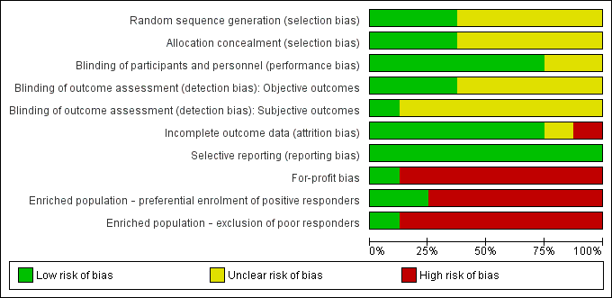
Risk of bias of included studies: review authors' judgements about each risk of bias item presented as percentages across all included studies
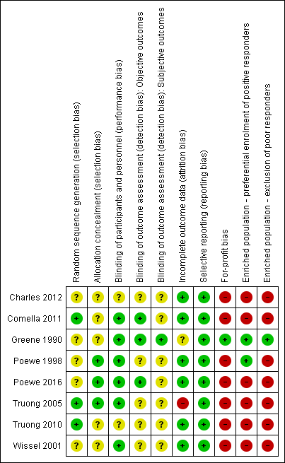
'Risk of bias' summary: review authors' judgements about each risk of bias item for each included study
Overall, we considered no studies to be at low risk of bias across all domains. However, the only two domains that we considered to represent a high risk of bias were the 'for‐profit bias' and 'enriched population' domains. It is noteworthy that all trials but one had a high risk of both 'for‐profit bias' and 'enriched population'.
Allocation
Three studies (Comella 2011; Truong 2005; Truong 2010) clearly described the process of random sequence generation; we assessed the other five studies to be at unclear risk of bias for this criterion. Poewe 1998, Poewe 2016, and Truong 2005 described an adequate allocation concealment process and we rated them as being at a low risk. All but one of the included trials reported a higher disease impairment at baseline in the control arm although it is unclear whether these differences are either statistically significant or clinically relevant. This leads us to assess the overall risk of selection bias across the included trials to be a serious cause for concern, despite the fact that overall no one trial is uniquely responsible for this assessment.
Blinding
We judged the blinding of participants and personnel involved in the trial to be at a low risk of bias in the majority of studies included in this review; three trials (Charles 2012; Poewe 2016; Truong 2010) did not describe enough information to allow for a clear judgment. We considered that only Greene 1990 had adequately blinded participants and investigators measuring both objective and subjective outcomes. For the assessment of objective outcomes, we also judged Comella 2011 to be at low risk. We considered that the remaining studies were at an unclear risk of bias across this domain. We judged Poewe 1998 at low risk of performance bias, but at an unclear risk of both elements of detection bias.
Incomplete outcome data
Six out of eight studies summarised the reasons for missing data and used appropriate statistical tools to deal with it, and we rated them at low risk of bias. Greene 1990 reported missing data across the study visits so that we assessed this study at unclear risk of attrition bias. Truong 2005 reported a large number of dropouts in both intervention arms, though this was asymmetrical, with over 60% of participants withdrawing from the placebo arm by week 4. For this reason we rated Truong 2005 at a high risk of attrition bias.
Selective reporting
We considered all studies to be at low risk for reporting bias.
Other potential sources of bias
For‐profit bias
All trials but one (Greene 1990) declared funding or supply of study vials from industry sources, and we rated them at a high risk of bias for funding and potential conflicts of interest.
Enriched population
Three studies (Charles 2012; Poewe 2016; Truong 2010) preferentially enrolled participants known to have previously responded to BtA treatment, and were at high risk of bias for enriched population. On the other hand, all studies except one (Greene 1990) excluded forms of cervical dystonia known to have a poorer clinical response to BtA injection, and were also considered to be at a high risk of bias for this domain.
Publication bias
We intended to use funnel plots to explore publication bias. However, due to the small number of included studies, the power of this analysis was considered to be inadequate (Sterne 2011).
Effects of interventions
The key results of this review can be found in summary of findings Table for the main comparison.
Preceding data analysis
See Dealing with missing data.
Poewe 1998, Poewe 2016, and Truong 2005 reported no SD for the primary outcome, so we opted to impute these values from other studies in this review (Wissel 2001; Truong 2010; Truong 2010, respectively), which used the same scale, time point and error measurement for assessing the same outcome.
Charles 2012 did not report the absolute numerical values for the primary outcome at week 4. Therefore, we opted to have two review authors independently extract these data from the graph provided in the report. Since the two review authors reported very similar values, we imputed these data (mean values from the two authors) and used them for analysis.
Poewe 1998 and Comella 2011 presented data separately for each BtA dose, reporting sample sizes, means and SD for each intervention group. In order to conduct pooled analyses we combined the BtA groups using a pooled SD formula for paired data.
Primary outcomes
Cervical dystonia‐specific improvement
The included trials assessed the primary outcome at either week 4 (Charles 2012; Comella 2011; Poewe 1998; Poewe 2016; Truong 2005; Truong 2010; Wissel 2001) or week 6 (Greene 1990) following initial injection of study medication. Most trials reported the primary efficacy outcome as the change from baseline measurement using validated scales, namely the CDSS (Charles 2012), the Tsui scale (Poewe 1998; Wissel 2001) and the TWSTRS (Comella 2011; Poewe 2016; Truong 2005; Truong 2010). Greene 1990 reported no objective efficacy measurements. The CDSS uses a protractor and wall chart to rate the severity of the head's deviation. The Tsui scale (range, 0 to 25) grades severity of postural deviance, acknowledging the presence of tremor and the pattern of movements; it does not assess disability, pain or other subjective symptoms. TWSTRS (range, 0 to 85) is composed of three subscales that grade severity (range, 0 to 35), disability (range, 0 to 30), and pain (range, 0 to 20). Tarsy 1997 demonstrated that Tsui and TWSTRS score reduction rates after botulinum toxin therapy correlate significantly with each other.
Seven trials (Charles 2012; Comella 2011; Poewe 1998; Poewe 2016; Truong 2005; Truong 2010; Wissel 2001; combined n = 833) contributed data for this outcome. Overall, treatment with BtA was associated with a moderate‐to‐large cervical dystonia‐specific improvement (SMD 0.70, 95% CI 0.52 to 0.89; I2 = 36%; moderate certainty in the evidence; Analysis 1.1).
Four trials (Comella 2011; Poewe 2016; Truong 2005; Truong 2010; combined n = 522) used the TWSTRS total score to assess cervical dystonia‐specific improvement. Overall, treatment with BtA was associated with a MD of 8.06 in TWSTRS total score compared to placebo (95% CI 6.08 to 10.05; I2 = 0%; Analysis 1.2), representing a 18.7% improvement compared to the baseline clinical status (43.55 TWSTRS combined baseline score).
Three trials (Comella 2011; Truong 2005; Truong 2010; combined n = 429) contributed data regarding the TWSTRS subscales. Overall, treatment with BtA was associated with a MD of 3.13 in the TWSTRS severity subscale (95% CI 2.15 to 4.11; I2 = 0%; Analysis 1.3), and 2.52 in the TWSTRS disability subscale (95% CI 1.72 to 3.31; I2 = 23%; Analysis 1.4).
For the Trial Sequential Analysis we could not use the results of the overall improvement, since these data were only available as SMD (Thorlund 2011). Thus, we opted to use the data from trials that used the TWSTRS scale.
The required information size was based on the event proportion or standard deviation in the control group; assumption of a plausible relative risk reduction (RRR) of 10%; a 5% risk of type I error; a 20% risk of type II error (power = 80%); and the observed heterogeneity of the meta‐analysis. We assumed a baseline TWSTRS of 42 points and an SD of 10. Given these constraints, the cumulative evidence overcame the heterogeneity‐adjusted required information size ‐ 180 participants. We are able to conclude that the cumulative evidence is adequately‐powered to demonstrate the 8.16 TWSTRS point difference at week 4 between BtA and placebo.
1.1. Overall improvement with high versus medium versus low dose of BtA subgroup analysis
We carried out a preplanned subgroup analysis to assess overall improvement according to the BtA dosages used (see Subgroup analysis and investigation of heterogeneity). Considering the current evidence behind the potency equivalence between BtA formulations, we assigned arbitrary thresholds for high, medium, and low doses of BtA. Two trials (Comella 2011; Poewe 1998; combined n = 193) contributed data to the high‐dose subgroup; six trials (Comella 2011; Poewe 1998; Poewe 2016; Truong 2005; Truong 2010; Wissel 2001; combined n = 545) contributed data to the medium‐dose subgroup; and one trial (Poewe 1998; n = 39) contributed data to the low‐dose subgroup.
One study (Charles 2012) reported a range of BtA injection doses that crossed the arbitrary dose limits we defined. We therefore did not include it in this subgroup meta‐analysis.
All three dosages were efficacious against placebo (high dose: SMD 1.08, 95% CI 0.53 to 1.63; I2 = 52%; medium dose: SMD 0.76, 95% CI 0.59 to 0.94; I2 = 0%; low dose: SMD 1.24, 95% CI 0.55 to 1.94), though we did not find a difference in efficacy between the subgroups (P = 0.25; Analysis 1.5).
1.2. Overall improvement with Botox versus Dysport versus Xeomin subgroup analysis
We carried out a preplanned subgroup analysis to assess overall improvement according to BtA formulation (see Subgroup analysis and investigation of heterogeneity). One trial (Charles 2012; n = 170) contributed data to the Botox subgroup; five trials (Poewe 1998; Poewe 2016; Truong 2005; Truong 2010; Wissel 2001; combined n = 430) contributed data to the Dysport subgroup; and one trial (Comella 2011; n = 233) contributed data to the Xeomin subgroup.
All three formulation were efficacious against placebo (Botox: SMD 0.38, 95% CI 0.08 to 0.69; Dysport: SMD 0.75, 95% CI 0.54 to 0.96; I2 = 8%; Xeomin: SMD 0.82, 95% CI 0.53 to 1.10), though we did not find a difference in efficacy between the subgroups (P = 0.08; Analysis 1.6).
1.3 Overall improvement with EMG‐guided versus non‐EMG‐guided injection subgroup analysis
We carried out a preplanned subgroup analysis to assess the comparative efficacy of BtA in trials that used EMG versus trials that did not use EMG (see Subgroup analysis and investigation of heterogeneity). Four trials (Comella 2011; Poewe 2016; Truong 2005; Truong 2010; combined n = 522) contributed data to the EMG‐guided subgroup; and three trials (Charles 2012; Poewe 1998; Wissel 2001; combined n = 311) contributed data to the non‐EMG‐guided subgroup.
It is important to note that all four trials that contributed data to the EMG‐guided subgroup left the use of EMG at the discretion of the investigator.
Both comparisons were efficacious against placebo (EMG‐guided: SMD 0.71, 95% CI 0.52 to 0.89; I2 = 0%; non‐EMG‐guided: SMD 0.79; 95% CI 0.27 to 1.31; I2 = 75%), though we did not find a difference in efficacy between the subgroups (P = 0.76; Analysis 1.7).
Adverse events
Seven trials (Charles 2012; Comella 2011; Poewe 1998; Poewe 2016; Truong 2005; Truong 2010; Wissel 2001: combined n = 952) contributed data for this outcome. Adverse events related to study treatment were reported in 55.3% of participants in the BtA groups, compared to 46.5% of participants in the placebo arms. Overall, treatment with BtA was associated with a 20% increase in the risk of adverse events, when compared with placebo (risk ratio (RR) 1.19; 95% CI 1.03 to 1.36; I2 = 16%; moderate certainty in the evidence; Analysis 1.8). The number needed to treat for an additional harmful outcome (NNTH) with a single BtA treatment was 9 (95% CI 5 to 31).
Adverse events were measured as the proportion of trial participants who experienced any adverse event during any point in follow‐up.
The required information size was based on the event proportion or standard deviation in the control group; assumption of a plausible relative risk reduction (RRR) of 10%; a 5% risk of type I error; a 20% risk of type II error (power = 80%); and the observed heterogeneity of the meta‐analysis. We assumed a control event rate of 46%. Given these constraints, the cumulative evidence overcame the heterogeneity‐adjusted required information size ‐ 892 participants. We are able to conclude that the cumulative evidence is adequately‐powered to demonstrate the 23% risk difference in adverse events between BtA and placebo.
2.1 Adverse events with high versus medium versus low dose of BtA subgroup analysis
We carried out a preplanned subgroup analysis to assess the risk of adverse events according to the BtA dosages used (see Subgroup analysis and investigation of heterogeneity). Considering the current evidence behind the potency equivalence between BtA formulations, we assigned arbitrary thresholds for low, medium, and high doses of BtA. Two trials (Comella 2011; Poewe 1998; combined n = 193) contributed data to the high‐dose subgroup; six trials (Comella 2011; Poewe 1998; Poewe 2016; Truong 2005; Truong 2010; Wissel 2001; combined n = 664) contributed data to the medium‐dose subgroup; and one trial (Poewe 1998; n = 39) contributed data to the low‐dose subgroup.
We did not find a difference in the risk of adverse events between the subgroups (P = 0.66; Analysis 1.9).
2.2 Adverse events with Botox versus Dysport versus Xeomin subgroup analysis
We carried out a preplanned subgroup analysis to assess the risk of adverse events according to BtA formulation (see Subgroup analysis and investigation of heterogeneity). One trial (Charles 2012; n = 170) contributed data to the Botox subgroup; five trials (Poewe 1998; Poewe 2016; Truong 2005; Truong 2010; Wissel 2001; n = 549) contributed data to the Dysport subgroup; and one trial (Comella 2011; n = 233) contributed data to the Xeomin subgroup.
Overall, we did not find a difference in the risk of adverse events between these subgroups (P = 0.34; Analysis 1.10).
2.3 Adverse events with EMG‐guided versus non‐EMG‐guided injection subgroup analysis
We carried out a preplanned subgroup analysis to assess the risk of adverse events in trials that used EMG versus trials that did not use EMG (see Subgroup analysis and investigation of heterogeneity). Four trials (Comella 2011; Poewe 2016; Truong 2005; Truong 2010; n = 640) contributed data to the EMG‐guided subgroup; and three trials (Charles 2012; Poewe 1998; Wissel 2001; n = 312) contributed data to the non‐EMG‐guided subgroup.
It is important to note that all four trials that contributed data to the EMG‐guided subgroup left the use of EMG at the discretion of the investigator.
Overall, we did not find a difference in the risk of adverse events between these subgroups (P = 0.52; Analysis 1.11).
2.4 Adverse events of special interest
Treatment with BtA was only associated with an increased risk of two adverse events of special interest, namely dysphagia (RR 3.04, 95% CI 1.68 to 5.50; I2 = 0%; moderate certainty in the evidence; Analysis 1.12), and diffuse weakness/tiredness (RR 1.78, 95% CI 1.08 to 2.94; I2 = 0%; moderate certainty in the evidence; Analysis 1.13). The NNTH with a single BtA treatment for dysphagia was 16 (95% CI 7 to 49) and that for diffuse weakness/tiredness was 21 (95% CI 9 to 208).
The remaining adverse events that were reported in the included trials included neck weakness (RR 3.23, 95% CI 0.95 to 10.91; I2 = 23%; Analysis 1.14), voice changes/hoarseness (RR 1.83, 95% CI 0.37 to 8.95; I2 = 26%; Analysis 1.15), sore throat/dry mouth (RR 1.66, 95% CI 0.78 to 3.51; I2 = 5%; Analysis 1.16), vertigo/dizziness (RR 1.47, 95% CI 0.38 to 5.73; I2=0%; Analysis 1.17), malaise/upper respiratory infection (RR 1.29, 95% CI 0.63 to 2.64; I2 = 45%; Analysis 1.18), injection site pain (RR 1.33, 95% CI 0.88 to 2.02; I2 = 0%; Analysis 1.19), and headache (RR 1.05, 95% CI 0.59 to 1.86; I2 = 0%; Analysis 1.20), though we did not find a difference between BtA and placebo regarding the risk of each one of these adverse events.
Secondary outcomes
Subjective evaluation of clinical status
The included trials assessed subjective evaluation of overall improvement by both physicians and participants between weeks 3 to 6 after BtA injection. They used four scales to evaluate the amount of improvement: the Patient Evaluation of Global Response (PEGR), the Global Assessment of Change (GAS), the visual analogue scale (VAS), and the Clinical Global Rating. PERG and GAS (range, ‐4 to +4) are similar scales ranging from "Very marked worsening" (‐4) to "Complete resolution of cervical dystonia symptoms" (+4). VAS (range, 0 mm to 100 mm) assesses the change from baseline in symptom severity, where 0 mm indicates "Much worse", 50 mm "No change", and 100 mm "Symptom‐free". The Clinical Global Rating is a scale taking into account 6 grades of efficacy (excellent, good, moderate, slight improvement, no change, condition worse) and 4 grades of adverse events (none, mild, moderate, extreme).
The trials measured subjective assessments using these validated scales as the change from baseline to weeks 3 to 6.
We could not combine data from three studies (Greene 1990; Poewe 2016; Truong 2010) into the meta‐analysis for this outcome. Greene 1990 reported no data for the control group, Poewe 2016 reported a change from baseline for both study groups but did not report a measure of dispersion, and Truong 2010 did not report the change from baseline.
3.1. Subjective assessment by clinicians
Four trials (Charles 2012; Comella 2011; Poewe 1998; Wissel 2001; n = 544) contributed data for this outcome. Treatment with BtA was associated with an increased likelihood of clinical improvement when compared to placebo as assessed by physicians between weeks 4 and 20 after drug injection (RR 1.91, CI 1.47 to 2.49; I2 = 28%; Analysis 1.21). We calculated an NNTB of 3 (95% CI 2 to 6) with a single BtA treatment session.
3.2. Subjective assessment by participants
Five trials (Charles 2012; Comella 2011; Poewe 1998; Truong 2005; Wissel 2001; n = 624) contributed data for this outcome. Treatment with BtA was associated with an increased likelihood of clinical improvement when compared to placebo as assessed by participants between weeks 4 and 20 after drug injection (RR 2.30, CI 1.83 to 2.90; I2 = 0%; moderate certainty in the evidence; Analysis 1.22). We calculated an NNTB of 3 (95% CI 2 to 5) with a single BtA treatment session.
Two trials (Poewe 1998; Truong 2005) considered only those participants reporting more than a half of improvement from baseline, and therefore these pooled results may underestimate the likelihood of participants reporting a subjective benefit with BtA treatment.
Pain relief
Six trials (Charles 2012; Comella 2011; Greene 1990; Truong 2005; Truong 2010; Wissel 2001; n = 722) contributed data to this outcome. They measured pain relief with validated pain scales as the change from baseline to weeks 3 to 6. Overall, treatment with BtA was associated with a moderately increased efficacy with regards to pain relief at weeks 4 to 6 (SMD 0.50, 95% CI 0.35 to 0.65; I2 = 0%; moderate certainty in the evidence; Analysis 1.26).
Three trials (Comella 2011; Truong 2005; Truong 2010; n = 429) contributed data measured on the TWSTRS pain subscale, and for this pooled result we found a MD of 2.11 TWSTRS points favouring participants allocated to BtA (95% CI 1.38 too 2.83; I2 = 0%; Analysis 1.27).
Health‐related quality of life
Three studies (Poewe 2016; Truong 2005; Truong 2010) assessed the impact of BtA on quality of life.
Poewe 2016 (n = 213) used the Cervical Dystonia Impact Profile (CDIP)‐58 scale (including eight subscales: head and neck symptoms, pain and discomfort, sleep, upper limb activities, walking, annoyance, mood and psychosocial functioning), and reported a significant improvement in total CDIP‐58 score (49.3 in BtA versus 59.4 in placebo; P < 0.0001), as well as in all eight CDIP‐58 subscales (P < 0.0003).
Truong 2005 (n = 80) and Truong 2010 (n = 116) reported an improvement from baseline to week 8 in the physical function domain of the SF‐36 (Truong 2005: odds ratio (OR) 1.60, P = 0.011; Truong 2010: MD 10.10, 95% CI 2.95 to 17.25; P = 0.018), but no benefit in social functioning when compared to placebo (Truong 2005: OR 0.30, 95% CI 0.23 to 0.82; P = 0.265; Truong 2010: MD 6.90, 95% CI 2.16 to 15.96; P = 0.125).
Tolerability
Adverse drug reactions (ADRs) can be a result of adverse events cause by the intervention (i.e. type A and/or type B ADRs), or lack of efficacy of the treatment (i.e. failure of therapy, a type F ADR) (Edwards 2000).
Four trials (Charles 2012; Comella 2011; Greene 1990; Truong 2010; n = 574) contributed data to this outcome. Overall, BtA was associated with a decreased likelihood of withdrawal from trials (RR 0.36, 95% CI 0.21 to 0.61; I2 = 0%; moderate certainty in the evidence; Analysis 1.30).
Three trials (Charles 2012; Comella 2011; Truong 2010; n = 519) contributed data regarding the withdrawals due to lack of efficacy. Overall, BtA was associated with a decreased likelihood of withdrawal from trials due to this reason (RR 0.30, 95% CI 0.17 to 0.53; I2 = 0%; Analysis 1.31).
Two trials (Comella 2011; Greene 1990; n = 288) contributed data regarding the withdrawals due to adverse events. Overall, we did not find a difference between BtA and placebo regarding withdrawals due to adverse events (RR 3.10; 95% CI 0.36 to 26.74; I2 = 0%; Analysis 1.32).
Duration of effect
We did not pool results for this outcome due to the lack of combinable data.
Most studies assessed duration of effect on participants who showed some level of response to the assigned study treatment, either BtA or placebo. Poewe 1998 reported a dose‐dependent duration of effect, with request for re‐injection occurring at week 8 in 39%, 50% and 94% of participants treated with high‐, medium‐ to low‐dose, and placebo, respectively.
Truong 2005 reported a mean duration of effect of 22.8 weeks (SD = 12.5; range 9 to 46) until recurrence of symptoms, while Truong 2010 reported a mean time to re‐treatment of 14.4 weeks (range 4 to 30).
Poewe 2016 evaluated response to BtA over five treatment cycles. It reports that the duration of treatment effect for treatment responders was more than 85 days from cycles 2 to 5, irrespective of the dose used.
Discussion
Summary of main results
This updated review included eight randomised, parallel‐designed trials, that enrolled 1010 people with cervical dystonia, of whom 70% had been previously treated with botulinum toxin for their condition.
As can be seen in the summary of findings Table for the main comparison, in comparison to placebo, BtA was probably more efficacious in reducing cervical dystonia‐associated impairment, including disease severity, disability, and associated pain. Treatment with BtA also increased the likelihood that participants and clinicians would detect any form of improvement. Uncertanity remains over the effect of BtA on other domains of people's quality of life, such as social functioning or mental health.
However, treatment with BtA increased the risk of experiencing an adverse event. In particular, BtA increased in the risk of two specific adverse events of special interest, namely dysphagia and diffuse weakness/tiredness. No fatalities or serious adverse events were considered related to BtA treatment in any trial. Finally, treatment with BtA slightly decreased the risk of withdrawal from the included clinical trials. Data for special subpopulations, such as children and pregnant women, were not available.
We found low to moderate statistical heterogeneity for most efficacy and safety outcome estimates.
BtA doses
All dosages were efficacious against placebo, but we found no clear‐cut evidence of a dose‐response gradient. It is, however, noteworthy that these trials were not dose‐response studies, they were not adequately powered to assess this question, and that we based this conclusion on arbitrarily defined dose‐subgroup analyses.
BtA formulations
Although none of the trials was designed or powered to evaluate the comparative utility of the three most widely used formulations of BtA (Botox (onabotulinumtoxinA), Dysport (abobotulinumtoxinA), and Xeomin (incobotulinumtoxinA)), we did not find differences between these subgroups in terms of overall efficacy or safety.
Use of EMG
Although none of the trials was designed to evaluate the comparative utility of injection technique with or without EMG, we did not find differences between trials that allowed and those that did not allow the use of EMG, in terms of overall efficacy or safety.
Duration of effect
Results from the few studies that addressed the duration of clinical effect of a treatment cycle in this review were not conclusive, with time to re‐treatment ranging greatly, from 1 to 11 months. We could not adequately evaluate long‐term duration of effect as all trials but one evaluated only a single treatment session.
Overall completeness and applicability of evidence
All included trials addressed the primary research question directly, using similar and validated assessment tools. However, they did not fully report data for all outcomes, and in some cases we could not pool results and compare them across studies. This limited the amount of data available and, consequently, the confidence in overall conclusions.
The participants included in the trials were not fully representative of the overall population of people with cervical dystonia. The effects of population enrichment and the moderate overall disease impairment (as assessed by the baseline TWSTRS scores) preclude definite conclusions concerning all people with this condition.
Four noteworthy factors challenge the implementation of the evidence in this review. Firstly, there was a considerably heterogeneous regional distribution, with most trials being conducted in North America and Europe. Differences in clinical practice, training of experts, and local guidelines in other regions of the world may present an obstacle to the application of the evidence here demonstrated. Secondly, sample size across included trials was relatively small and many subgroup analyses addressing clinically relevant questions for the main outcomes were underpowered. More studies are needed to provide robust evidence for these questions. Thirdly, the use of enriched populations in clinical trials limits applicability of results into clinical practice, as complex and potentially poorer responders are usually excluded in these trials. The fact that such individuals are common in clinical practice further complicates issues of generalisation. Fourthly, it is common for people with cervical dystonia to have concomitant medications for their condition, such as muscle relaxants and benzodiazepines. Reasonably, participants in trials are required to be on a stable dose of these medications for many weeks to avoid confounding factors. As a result, little is known at present about the impact of these drug regimens with regard to implementation of the evidence in this review.
Quality of the evidence
See Characteristics of included studies, 'Risk of bias' tables, 'Risk of bias' summary tables (Figure 2; Figure 3), and summary of findings Table for the main comparison.
We considered all included trials but one to be at a high risk for both 'for‐profit bias' and having an enriched population. Only three of the included studies adequately described the randomisation or allocation methods, or both, with the remaining trials being assessed at an unclear risk of bias for these items. We considered most studies to be appropriately blinded in general. However, only a single trial provided a satisfactory description of blinding of objective outcome assessment, and we considered all but one possibly biased regarding subjective outcome assessment, as most studies predominantly enrolled participants with previous exposure to botulinum toxin. This represents a major methodological limitation that may have resulted in a biased assessment of the intervention effect, particularly with regards to subjective outcomes, namely pain assessment, subjective assessment by participants and clinicians, and quality of life assessments, which are highly susceptible to biased estimations.
We could not compare some outcomes across studies, as some studies did not report relevant data. We were unable to impute values for missing data due to imbalances between baseline characteristics of the participants and incomplete description of the variables, further reducing the amount of combinable data, and therefore our confidence in the results.
The included trials each enrolled between 55 and 233 participants, and although individually some of these trials were underpowered, the pooling of the trials permitted an adequate sample size for the majority of efficacy outcomes.
Taken together, as can be seen in summary of findings Table for the main comparison, we consider that there is moderate certainty in the evidence that a single treatment session of BtA improves cervical dystonia‐associated impairment in certain types of cervical dystonia, including severity, disability, and pain. The certainty in the evidence supporting the higher occurrence of any adverse event, as well diffuse weakness/tiredness and dysphagia is moderate. The certainty in the evidence assessing the change in subjective evaluation of clinical status evaluated by participants is moderate. Finally, we have moderate certainty in the evidence that treatment with BtA increases the likelihood of participants not dropping out of clinical trials.
Potential biases in the review process
Although we followed the methods recommended by Cochrane in order to minimise bias in the review process, certain areas do deserve attention. In particular, we have not searched clinical trials registries. Although this opens the current review to the potential bias of having missed trials, we consider this possibility highly unlikely because we have extensively contacted other experts in this field and US and European trials in this area are well‐known.
An additional bias was that we could not obtain data for all outcomes in the included trials. A further limitation of this review is the small number of participants contributing data to each outcome, although Trial Sequential Analysis showed that both the primary efficacy and safety outcomes were adequately powered to demonstrate the difference that we observed.
Agreements and disagreements with other studies or reviews
Overall, the results of this updated review are in agreement with the conclusions of earlier versions (Costa 2005). The current clinical practice guidelines of the American Academy of Neurology and the European Academy of Neurology agree that BtA is "established as safe and effective for cervical dystonia treatment" (Simpson 2016) and that it is considered an "effective and safe treatment for cervical dystonia" (Albanese 2011). We now conclude that no claims can be made regarding a clear dose‐response relationship for efficacy outcomes. On the other hand, a clear relationship exists for the increased risk of treatment‐related adverse events of special interest, namely dysphagia and diffuse weakness/tiredness.

Study flow diagram

Risk of bias of included studies: review authors' judgements about each risk of bias item presented as percentages across all included studies

'Risk of bias' summary: review authors' judgements about each risk of bias item for each included study
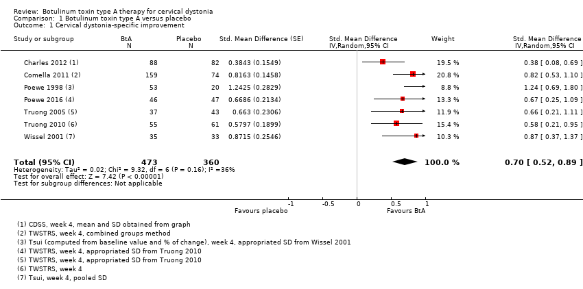
Comparison 1 Botulinum toxin type A versus placebo, Outcome 1 Cervical dystonia‐specific improvement.

Comparison 1 Botulinum toxin type A versus placebo, Outcome 2 Cervical dystonia‐specific improvement ‐ TWSTRS subgroup analysis.
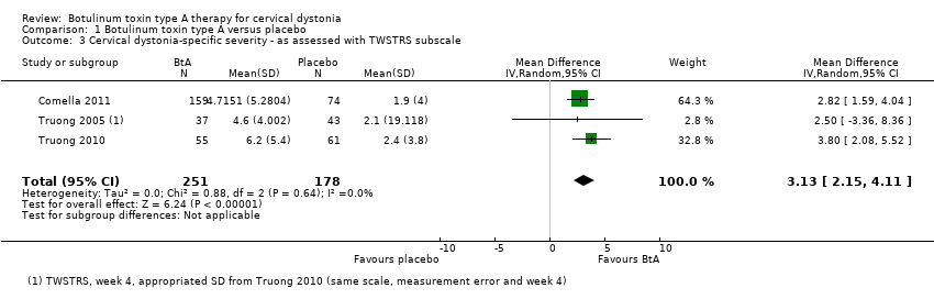
Comparison 1 Botulinum toxin type A versus placebo, Outcome 3 Cervical dystonia‐specific severity ‐ as assessed with TWSTRS subscale.

Comparison 1 Botulinum toxin type A versus placebo, Outcome 4 Cervical dystonia‐specific disability ‐ as assessed with TWSTRS subscale.

Comparison 1 Botulinum toxin type A versus placebo, Outcome 5 Cervical dystonia‐specific improvement ‐ doses subgroup analysis.
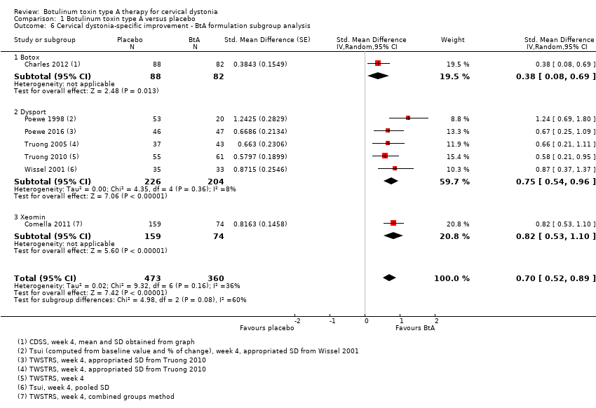
Comparison 1 Botulinum toxin type A versus placebo, Outcome 6 Cervical dystonia‐specific improvement ‐ BtA formulation subgroup analysis.
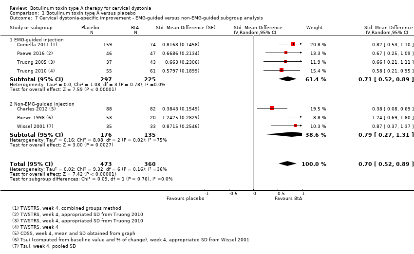
Comparison 1 Botulinum toxin type A versus placebo, Outcome 7 Cervical dystonia‐specific improvement ‐ EMG‐guided versus non‐EMG‐guided subgroup analysis.

Comparison 1 Botulinum toxin type A versus placebo, Outcome 8 Adverse events.

Comparison 1 Botulinum toxin type A versus placebo, Outcome 9 Adverse events ‐ doses subgroup analysis.

Comparison 1 Botulinum toxin type A versus placebo, Outcome 10 Adverse events ‐ BtA formulation subgroup analysis.
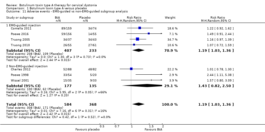
Comparison 1 Botulinum toxin type A versus placebo, Outcome 11 Adverse events ‐ EMG‐guided vs non‐EMG‐guided subgroup analysis.
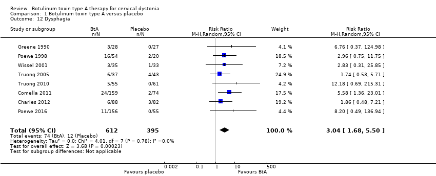
Comparison 1 Botulinum toxin type A versus placebo, Outcome 12 Dysphagia.
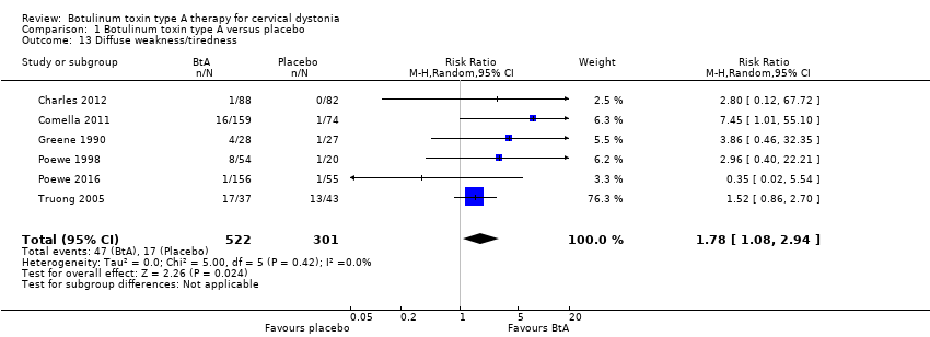
Comparison 1 Botulinum toxin type A versus placebo, Outcome 13 Diffuse weakness/tiredness.

Comparison 1 Botulinum toxin type A versus placebo, Outcome 14 Neck weakness.
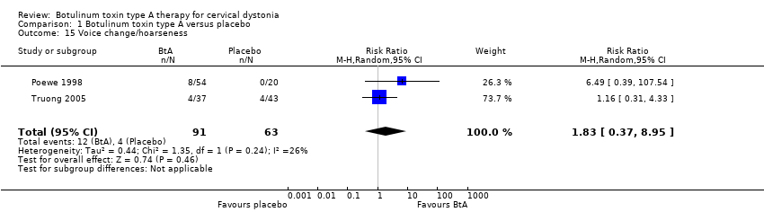
Comparison 1 Botulinum toxin type A versus placebo, Outcome 15 Voice change/hoarseness.
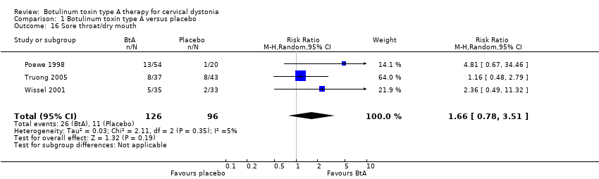
Comparison 1 Botulinum toxin type A versus placebo, Outcome 16 Sore throat/dry mouth.
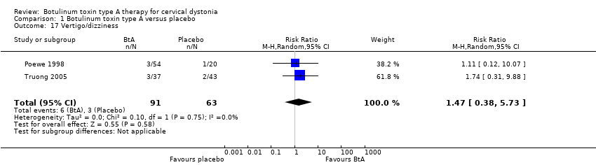
Comparison 1 Botulinum toxin type A versus placebo, Outcome 17 Vertigo/dizziness.
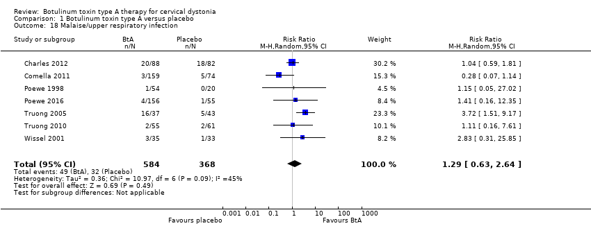
Comparison 1 Botulinum toxin type A versus placebo, Outcome 18 Malaise/upper respiratory infection.

Comparison 1 Botulinum toxin type A versus placebo, Outcome 19 Local pain (injection site).
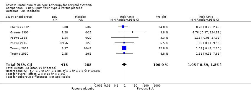
Comparison 1 Botulinum toxin type A versus placebo, Outcome 20 Headache.

Comparison 1 Botulinum toxin type A versus placebo, Outcome 21 Any improvement by subjective clinician assessment.

Comparison 1 Botulinum toxin type A versus placebo, Outcome 22 Any improvement by subjective participant assessment.

Comparison 1 Botulinum toxin type A versus placebo, Outcome 23 Any improvement by subjective participant assessment ‐ doses subgroup analysis.

Comparison 1 Botulinum toxin type A versus placebo, Outcome 24 Any improvement by subjective participant assessment ‐ BtA formulation subgroup analysis.

Comparison 1 Botulinum toxin type A versus placebo, Outcome 25 Any improvement by subjective participant assessment ‐ EMG guided vs non‐EMG‐guided subgroup analysis.
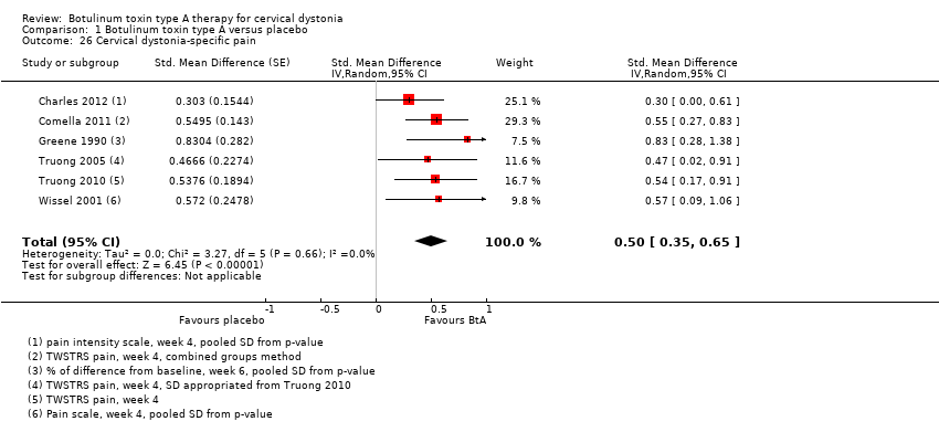
Comparison 1 Botulinum toxin type A versus placebo, Outcome 26 Cervical dystonia‐specific pain.

Comparison 1 Botulinum toxin type A versus placebo, Outcome 27 Cervical dystonia‐specific pain ‐ TWSTRS pain subscale subgroup analysis.

Comparison 1 Botulinum toxin type A versus placebo, Outcome 28 Cervical dystonia‐specific pain ‐ BtA formulation subgroup analysis.

Comparison 1 Botulinum toxin type A versus placebo, Outcome 29 Cervical dystonia‐specific pain ‐ EMG‐guided vs non‐EMG‐guided subgroup analysis.

Comparison 1 Botulinum toxin type A versus placebo, Outcome 30 Tolerability ‐ withdrawals.

Comparison 1 Botulinum toxin type A versus placebo, Outcome 31 Tolerability ‐ withdrawals due lack of efficacy subgroup analysis.

Comparison 1 Botulinum toxin type A versus placebo, Outcome 32 Tolerability ‐ withdrawals due to adverse events subgroup analysis.
| Botulinum toxin type A compared to placebo for cervical dystonia | ||||||
| Patient or population: adults with cervical dystonia | ||||||
| Outcomes | Relative effect | Anticipated absolute effects* (95% CI) | Certainty in the evidence | What happens | ||
| With placebo | With botulinum toxin type A | Difference | ||||
| Cervical dystonia‐specific impairment Assessed 3 to 6 weeks post‐injection using TWSTRS total score | ‐ | 3.9 TWSTRS units decrease | 12.45 TWSTRS units decrease | The mean change from baseline was 8.06 TWSTRS units higher (6.08 higher to 10.05 higher) in the BtA group compared to the placebo group | ⊕⊕⊕⊝ | BtA treatment probably improves cervical dystonia‐specific impairment |
| Adverse events Assessed at any point during follow‐up | RR 1.19 | 46.5% | 55.3% | 8.8% more | ⊕⊕⊕⊝ | BtA treatment probably increases the risk of adverse events |
| Subjective participant assessment Assessed 3 to 6 weeks post‐injection | RR 2.30 | 24.2% | 55.7% | 31.5% more | ⊕⊕⊕⊝ | BtA treatment probably increases the likelihood that patients will detect any form of improvement |
| Pain relief Assessed 3 to 6 weeks post‐injection using TWSTRS pain subscore | ‐b | ‐b | ‐b | The mean change from baseline was 2.11 TWSTRS units higher (1.38 higher to 2.83 higher) in the BtA group compared to the placebo group | ⊕⊕⊕⊝ | BtA treatment probably improves cervical dystonia‐related pain |
| Tolerability Assessed at any point during follow‐up | RR 0.38 | 20.5% | 7.8% (4.7 to 12.7) | 12.7% fewer (15.8 to 7.8) | ⊕⊕⊕⊝ | BtA treatment probably slightly decreases the risk of withdrawal of clinical trials |
| Dysphagia Assessed at any point during follow‐up | RR 3.04 | 3.0% | 9.2% | 6.2% more | ⊕⊕⊕⊝ | BtA treatment probably increases the risk of dysphagia |
| Diffuse weakness/tiredness Assessed at any point during follow‐up | RR 1.78 | 5.6% | 10.1% | 4.4% more | ⊕⊕⊕⊝ | BtA treatment probably increases the risk of diffuse weakness/tiredness |
| *The risk in the intervention group (and its 95% confidence interval) is based on the assumed risk in the comparison group and the relative effect of the intervention (and its 95% CI). | ||||||
| GRADE Working Group grades of evidence | ||||||
| a Downgraded one level due to serious study limitations, namely concerns with randomisation procedures and other biases such as 'for‐profit' bias. | ||||||
| Term | Definition |
| BtA‐non‐responsive | People who do not experience the expected benefit from treatment with botulinum toxin type A |
| Cervical dystonia or spasmodic torticollis | A common movement disorder in which people have abnormal movements or postures of the head and neck that they cannot control. It is frequently accompanied by social embarrassment and pain. |
| Chemodenervation | The process by which botulinum toxin causes muscular paralysis. Although all the anatomical elements necessary for muscular control are intact (i.e. nerve, synapse and muscle), there is a chemical process that disables the transmission of the electrical signal from the nerve to the muscle. |
| Dysphagia | A discomfort or difficulty when swallowing. |
| Electromyography | An examination that displays the electrical activity of muscles using pieces of metal attached to the skin or inserted into the muscle. |
| Non‐naive | People who have been treated in the past with botulinum toxin. |
| Voluntary action | Movements that people are able to control, start and stop when they want to. |
| Study ID | Number of participants | Total dropouts | Age mean, range (years) | Baseline CD impairment BtA/placebo | % participants naive to Bt | BtA formulation | Total dose per participant | EMG‐guidance | Study duration (weeks) |
| Charles 2012 | 170 | 35 (11 in BtA) | 55, 31‐76 | 9.2/9.3 (CDSS) | 0 | Botox (OnaBtA) | 236 | No | 10 |
| Comella 2011 | 233 | 14 (8 in BtA) | 53 | 42.4/41.8 (TWSTRS) | 39 | Xeomin (IncoBtA) | 120 or 240 | At discretion | 20 |
| Greene 1990 | 55 | 3 (3 in BtA) | 50 | 21% severe/ 41% severe | 100 | Botox (OnaBtA) | 150 to 165 | No | 12 |
| Poewe 1998 | 75 | 2 (2 in BtA) | 47, 26‐82 | 13.9/14.4 (Tsui scale) | 100 | Dysport (AboBtA) | 250, 500 or 1000 | No | 8 |
| Poewe 2016 | 213 | N/A | 49 | 46/47 (TWSTRS) | 10 | Dysport (AboBtA) | 500 | N/A | 12 |
| Truong 2005 | 80 | 56 (22 in BtA) | 54, 27‐78 | 45.1/46.2 (TWSTRS) | 26 | Dysport (AboBtA) | 500 | At discretion | 20 |
| Truong 2010 | 116 | 33 (10 in BtA) | 53, 20‐79 | 43.8/45.8 (TWSTRS) | 17 | Dysport (AboBtA) | 500 | At discretion | 12 |
| Wissel 2001 | 68 | 0 | 48, 18‐75 | 11.1/11.5 (Tsui scale) | 31 | Dysport (AboBtA) | 500 | No | 16 |
| Bt: botulinum toxin; CD: cervical dystonia; CDSS: Cervical Dystonia Severity Scale; EMG: electromyography; TWSTRS: Toronto Western Spasmodic Torticollis Rating Scale | |||||||||
| Outcome or subgroup title | No. of studies | No. of participants | Statistical method | Effect size |
| 1 Cervical dystonia‐specific improvement Show forest plot | 7 | 833 | Std. Mean Difference (Random, 95% CI) | 0.70 [0.52, 0.89] |
| 2 Cervical dystonia‐specific improvement ‐ TWSTRS subgroup analysis Show forest plot | 4 | 522 | Mean Difference (IV, Random, 95% CI) | 8.06 [6.08, 10.05] |
| 3 Cervical dystonia‐specific severity ‐ as assessed with TWSTRS subscale Show forest plot | 3 | 429 | Mean Difference (IV, Random, 95% CI) | 3.13 [2.15, 4.11] |
| 4 Cervical dystonia‐specific disability ‐ as assessed with TWSTRS subscale Show forest plot | 3 | 429 | Mean Difference (IV, Random, 95% CI) | 2.52 [1.72, 3.31] |
| 5 Cervical dystonia‐specific improvement ‐ doses subgroup analysis Show forest plot | 6 | 777 | Std. Mean Difference (Random, 95% CI) | 0.84 [0.68, 1.00] |
| 5.1 Low dose | 1 | 39 | Std. Mean Difference (Random, 95% CI) | 1.24 [0.55, 1.94] |
| 5.2 Medium dose | 6 | 545 | Std. Mean Difference (Random, 95% CI) | 0.76 [0.59, 0.94] |
| 5.3 High dose | 2 | 193 | Std. Mean Difference (Random, 95% CI) | 1.08 [0.53, 1.63] |
| 6 Cervical dystonia‐specific improvement ‐ BtA formulation subgroup analysis Show forest plot | 7 | 833 | Std. Mean Difference (Random, 95% CI) | 0.70 [0.52, 0.89] |
| 6.1 Botox | 1 | 170 | Std. Mean Difference (Random, 95% CI) | 0.38 [0.08, 0.69] |
| 6.2 Dysport | 5 | 430 | Std. Mean Difference (Random, 95% CI) | 0.75 [0.54, 0.96] |
| 6.3 Xeomin | 1 | 233 | Std. Mean Difference (Random, 95% CI) | 0.82 [0.53, 1.10] |
| 7 Cervical dystonia‐specific improvement ‐ EMG‐guided versus non‐EMG‐guided subgroup analysis Show forest plot | 7 | 833 | Std. Mean Difference (Random, 95% CI) | 0.70 [0.52, 0.89] |
| 7.1 EMG‐guided injection | 4 | 522 | Std. Mean Difference (Random, 95% CI) | 0.71 [0.52, 0.89] |
| 7.2 Non‐EMG‐guided injection | 3 | 311 | Std. Mean Difference (Random, 95% CI) | 0.79 [0.27, 1.31] |
| 8 Adverse events Show forest plot | 7 | 952 | Risk Ratio (M‐H, Random, 95% CI) | 1.19 [1.03, 1.36] |
| 9 Adverse events ‐ doses subgroup analysis Show forest plot | 6 | Risk Ratio (M‐H, Random, 95% CI) | Subtotals only | |
| 9.1 Low dose | 1 | 39 | Risk Ratio (M‐H, Random, 95% CI) | 1.47 [0.56, 3.85] |
| 9.2 Medium dose | 6 | 664 | Risk Ratio (M‐H, Random, 95% CI) | 1.23 [1.06, 1.44] |
| 9.3 High dose | 2 | 193 | Risk Ratio (M‐H, Random, 95% CI) | 1.90 [0.72, 5.02] |
| 10 Adverse events ‐ BtA formulation subgroup analysis Show forest plot | 7 | 952 | Risk Ratio (M‐H, Random, 95% CI) | 1.19 [1.03, 1.36] |
| 10.1 Botox | 1 | 170 | Risk Ratio (M‐H, Random, 95% CI) | 1.01 [0.78, 1.30] |
| 10.2 Dysport | 5 | 549 | Risk Ratio (M‐H, Random, 95% CI) | 1.30 [1.02, 1.66] |
| 10.3 Xeomin | 1 | 233 | Risk Ratio (M‐H, Random, 95% CI) | 1.22 [0.92, 1.62] |
| 11 Adverse events ‐ EMG‐guided vs non‐EMG‐guided subgroup analysis Show forest plot | 7 | 952 | Risk Ratio (M‐H, Random, 95% CI) | 1.19 [1.03, 1.36] |
| 11.1 EMG‐guided injection | 4 | 640 | Risk Ratio (M‐H, Random, 95% CI) | 1.19 [1.03, 1.36] |
| 11.2 Non‐EMG‐guided injection | 3 | 312 | Risk Ratio (M‐H, Random, 95% CI) | 1.43 [0.82, 2.50] |
| 12 Dysphagia Show forest plot | 8 | 1007 | Risk Ratio (M‐H, Random, 95% CI) | 3.04 [1.68, 5.50] |
| 13 Diffuse weakness/tiredness Show forest plot | 6 | 823 | Risk Ratio (M‐H, Random, 95% CI) | 1.78 [1.08, 2.94] |
| 14 Neck weakness Show forest plot | 4 | 277 | Risk Ratio (M‐H, Random, 95% CI) | 3.23 [0.95, 10.91] |
| 15 Voice change/hoarseness Show forest plot | 2 | 154 | Risk Ratio (M‐H, Random, 95% CI) | 1.83 [0.37, 8.95] |
| 16 Sore throat/dry mouth Show forest plot | 3 | 222 | Risk Ratio (M‐H, Random, 95% CI) | 1.66 [0.78, 3.51] |
| 17 Vertigo/dizziness Show forest plot | 2 | 154 | Risk Ratio (M‐H, Random, 95% CI) | 1.47 [0.38, 5.73] |
| 18 Malaise/upper respiratory infection Show forest plot | 7 | 952 | Risk Ratio (M‐H, Random, 95% CI) | 1.29 [0.63, 2.64] |
| 19 Local pain (injection site) Show forest plot | 7 | 837 | Risk Ratio (M‐H, Random, 95% CI) | 1.33 [0.88, 2.02] |
| 20 Headache Show forest plot | 6 | 706 | Risk Ratio (M‐H, Random, 95% CI) | 1.05 [0.59, 1.86] |
| 21 Any improvement by subjective clinician assessment Show forest plot | 4 | 544 | Risk Ratio (M‐H, Random, 95% CI) | 1.91 [1.47, 2.49] |
| 22 Any improvement by subjective participant assessment Show forest plot | 5 | 624 | Risk Ratio (M‐H, Random, 95% CI) | 2.30 [1.83, 2.90] |
| 23 Any improvement by subjective participant assessment ‐ doses subgroup analysis Show forest plot | 4 | Risk Ratio (M‐H, Random, 95% CI) | Subtotals only | |
| 23.1 Low dose | 1 | 39 | Risk Ratio (M‐H, Random, 95% CI) | 1.58 [0.30, 8.43] |
| 23.2 Medium dose | 4 | 336 | Risk Ratio (M‐H, Random, 95% CI) | 2.44 [1.82, 3.25] |
| 23.3 High dose | 2 | 193 | Risk Ratio (M‐H, Random, 95% CI) | 3.39 [2.16, 5.33] |
| 24 Any improvement by subjective participant assessment ‐ BtA formulation subgroup analysis Show forest plot | 5 | Risk Ratio (M‐H, Random, 95% CI) | Subtotals only | |
| 24.1 Botox | 1 | 170 | Risk Ratio (M‐H, Random, 95% CI) | 1.99 [1.34, 2.94] |
| 24.2 Dysport | 3 | 221 | Risk Ratio (M‐H, Random, 95% CI) | 2.13 [1.49, 3.04] |
| 24.3 Xeomin | 1 | 233 | Risk Ratio (M‐H, Random, 95% CI) | 3.23 [2.03, 5.14] |
| 25 Any improvement by subjective participant assessment ‐ EMG guided vs non‐EMG‐guided subgroup analysis Show forest plot | 5 | Risk Ratio (M‐H, Random, 95% CI) | Subtotals only | |
| 25.1 EMG‐guided injection | 2 | 313 | Risk Ratio (M‐H, Random, 95% CI) | 2.97 [1.99, 4.43] |
| 25.2 Non‐EMG‐guided injection | 3 | 311 | Risk Ratio (M‐H, Random, 95% CI) | 2.03 [1.53, 2.69] |
| 26 Cervical dystonia‐specific pain Show forest plot | 6 | Std. Mean Difference (Random, 95% CI) | 0.50 [0.35, 0.65] | |
| 27 Cervical dystonia‐specific pain ‐ TWSTRS pain subscale subgroup analysis Show forest plot | 3 | Mean Difference (Random, 95% CI) | 2.11 [1.38, 2.83] | |
| 28 Cervical dystonia‐specific pain ‐ BtA formulation subgroup analysis Show forest plot | 6 | Std. Mean Difference (Random, 95% CI) | 0.50 [0.35, 0.65] | |
| 28.1 Botox | 2 | Std. Mean Difference (Random, 95% CI) | 0.51 [0.01, 1.02] | |
| 28.2 Dysport | 3 | Std. Mean Difference (Random, 95% CI) | 0.52 [0.28, 0.77] | |
| 28.3 Xeomin | 1 | Std. Mean Difference (Random, 95% CI) | 0.55 [0.27, 0.83] | |
| 29 Cervical dystonia‐specific pain ‐ EMG‐guided vs non‐EMG‐guided subgroup analysis Show forest plot | 6 | 654 | Std. Mean Difference (Random, 95% CI) | 0.50 [0.35, 0.65] |
| 29.1 EMG‐guided injection | 3 | 429 | Std. Mean Difference (Random, 95% CI) | 0.53 [0.33, 0.73] |
| 29.2 Non‐EMG‐guided injection | 3 | 225 | Std. Mean Difference (Random, 95% CI) | 0.50 [0.20, 0.80] |
| 30 Tolerability ‐ withdrawals Show forest plot | 4 | 574 | Risk Ratio (IV, Random, 95% CI) | 0.38 [0.23, 0.62] |
| 31 Tolerability ‐ withdrawals due lack of efficacy subgroup analysis Show forest plot | 3 | 519 | Risk Ratio (IV, Random, 95% CI) | 0.30 [0.17, 0.53] |
| 32 Tolerability ‐ withdrawals due to adverse events subgroup analysis Show forest plot | 2 | 288 | Risk Ratio (IV, Random, 95% CI) | 3.10 [0.36, 26.74] |