Resonancia magnética versus tomografía computarizada para la detección de lesiones vasculares agudas en pacientes que presentan síntomas de accidente cerebrovascular
Referencias
Referencias de los estudios incluidos en esta revisión
Referencias de los estudios excluidos de esta revisión
Referencias adicionales
Referencias de otras versiones publicadas de esta revisión
Characteristics of studies
Characteristics of included studies [ordered by study ID]
Ir a:
| Clinical features and settings | Patients with suspected acute ischaemic stroke in the middle cerebral artery territory who were studied with both DWI and CT within 6 hours of symptom onset | |
| Participants | 17 patients (53% men) presenting with stroke symptoms | |
| Study design | Prospective | |
| Target condition and reference standard(s) | Ischaemic stroke | |
| Index and comparator tests | DWI versus CT | |
| Follow‐up | T2‐weighted imaging performed at 90 days | |
| Notes | None of the patients were treated with thrombolysis | |
| Table of Methodological Quality | ||
| Item | Authors' judgement | Description |
| Representative spectrum? | No | Selected sample of patients |
| Acceptable reference standard? | Yes | Standard clinical criteria and imaging follow up (T2‐WI) |
| Acceptable delay between tests? | Yes | Mean time between index test (DWI) and comparator (CT) = 1 hour |
| Partial verification avoided? | Yes | All patients were verified by the reference criteria |
| Differential verification avoided? | Yes | All patients were verified by the same reference standard |
| Incorporation avoided? | Yes | Final diagnosis of acute stroke did not include the acute DW images and CT scans |
| Reference standard results blinded? | Unclear | Unclear whether the follow up T2‐W images were read blind to the acute images |
| Index test results blinded? | Yes | Acute CT and DWI images were read separately by 2 neuroradiologists who were blinded to patients' clinical data and results of the other imaging study |
| Relevant clinical information? | Yes | No clinical information was provided to the clinicians who interpreted CT and DW images |
| Uninterpretable results reported? | Yes | 1 patient could not tolerate MR |
| Withdrawals explained? | Yes | No withdrawals |
| Prospective study? | Yes | Prospective recruitment of consecutive patients |
| Expertise of imaging tests readers reported? | Yes | 2 neuroradiologists |
| Sequence of tests determined at random? | No | The order of tests was not randomised |
| Scans read blind to clinical information? | Yes | The neuroradiologists who interpreted CT and DWI results were blinded to patients' clinical details |
| Clinical features and settings | Patients with suspected acute ischaemic stroke who underwent imaging within 12 hours of symptom onset | |
| Participants | 15 stroke patients (40% men) | |
| Study design | Prospective | |
| Target condition and reference standard(s) | Acute ischaemic stroke | |
| Index and comparator tests | DWI versus CT | |
| Follow‐up | CT at 8 days | |
| Notes | Haemorrhage excluded | |
| Table of Methodological Quality | ||
| Item | Authors' judgement | Description |
| Representative spectrum? | No | Selected sample of patients |
| Acceptable reference standard? | Unclear | Probably standard clinical criteria and imaging follow up |
| Acceptable delay between tests? | Yes | Time between CT and DWI = 1 hour |
| Partial verification avoided? | Yes | All patients were verified by the reference criteria |
| Differential verification avoided? | Yes | All patients were verified by the same reference criteria |
| Incorporation avoided? | Yes | Final diagnosis did not include the results of acute CT and DWI |
| Reference standard results blinded? | Unclear | Not clearly reported |
| Index test results blinded? | Unclear | Unclear whether the acute CT and DW images were read blind to patients clinical details and final diagnosis |
| Relevant clinical information? | Unclear | Unclear whether the clinicians who interpreted the CT and DW images were provided with patients' clinical details |
| Uninterpretable results reported? | Unclear | Unclear whether patients with uninterpretable results were excluded |
| Withdrawals explained? | Yes | No withdrawals |
| Prospective study? | Yes | Prospective recruitment of consecutive patients |
| Expertise of imaging tests readers reported? | No | Not reported |
| Sequence of tests determined at random? | No | The order of tests was not randomised |
| Scans read blind to clinical information? | Unclear | Unclear whether the clinicians who interpreted CT and DW images were blinded to patients' clinical information |
| Clinical features and settings | Patients with suspected acute stroke who underwent both DWI and CT within 3 hours of symptom onset | |
| Participants | 90 patients presenting with stroke symptoms | |
| Study design | Prospective | |
| Target condition and reference standard(s) | Acute stroke | |
| Index and comparator tests | DWI versus CT for detection of ischaemic stroke | |
| Follow‐up | Imaging | |
| Notes | None of the patients were treated with thrombolysis | |
| Table of Methodological Quality | ||
| Item | Authors' judgement | Description |
| Representative spectrum? | Yes | A consecutive series of patients referred to the hospital's stroke team because of suspicion of acute stroke; irrespective of time from onset, symptom severity, or ultimate clinical diagnosis |
| Acceptable reference standard? | Yes | All available clinical information and follow up brain imaging |
| Acceptable delay between tests? | Unclear | Time between DWI and CT for detection of ischaemic stroke: 120 minutes |
| Partial verification avoided? | Yes | All patients were verified by the reference criteria |
| Differential verification avoided? | Unclear | Unclear whether all patients were verified by the same reference criteria |
| Incorporation avoided? | No | Final diagnosis included both acute and follow‐up brain images |
| Reference standard results blinded? | No | Follow‐up images were not read blind to the findings of acute images |
| Index test results blinded? | Yes | Acute images were read blind to patients clinical details and final diagnosis |
| Relevant clinical information? | Yes | No clinical information were provided to the clinicians who interpret CT and DW images |
| Uninterpretable results reported? | No | Uninterpretable brain images were excluded |
| Withdrawals explained? | Yes | No withdrawals |
| Prospective study? | Yes | Prospective recruitment of a series of consecutive patients |
| Expertise of imaging tests readers reported? | Yes | Images were analysed by 2 expert neuroradiologists and 2 expert stroke neurologists |
| Sequence of tests determined at random? | No | The order of tests was not randomised |
| Scans read blind to clinical information? | Yes | The clinicians who read the acute images were blinded to patients' clinical information and final diagnosis |
| Clinical features and settings | Patients with suspected ischaemic stroke and with a negative or inconclusive CT scan and for whom MRI was deemed essential for establishing proper management | |
| Participants | 22 patients (55% men) with acute stroke | |
| Study design | Retrospective | |
| Target condition and reference standard(s) | Acute ischaemic stroke | |
| Index and comparator tests | DWI versus CT | |
| Follow‐up | Clinical assessment and imaging | |
| Notes | Haemorrhage excluded | |
| Table of Methodological Quality | ||
| Item | Authors' judgement | Description |
| Representative spectrum? | No | Selected sample |
| Acceptable reference standard? | Yes | Clinical criteria and imaging follow up (CT or MRI) |
| Acceptable delay between tests? | Yes | Mean time between CT and DWI = 4.2 hours |
| Partial verification avoided? | Yes | All patients were verified by the reference criteria |
| Differential verification avoided? | Yes | All patients were verified by the same reference criteria |
| Incorporation avoided? | Yes | Final diagnosis did not include the acute CT and DW images |
| Reference standard results blinded? | Unclear | Unclear whether the physicians who confirmed the final diagnosis were aware of the acute CT and DWI findings |
| Index test results blinded? | Unclear | The neuroradiologists who interpreted the acute CT and DW images were not blind to patients' clinical history but they were reported to be blinded to final diagnosis |
| Relevant clinical information? | No | A brief description of patients' clinical symptoms was provided to the neuroradiologists who interpreted CT and DW images |
| Uninterpretable results reported? | Yes | No uninterpretable results |
| Withdrawals explained? | Yes | No withdrawals |
| Prospective study? | No | Retrospective study |
| Expertise of imaging tests readers reported? | Yes | Images were reviewed by a neuroradiology fellow and a staff neuroradiologist |
| Sequence of tests determined at random? | No | The order of tests was not randomised |
| Scans read blind to clinical information? | No | The 2 neuroradiologists who reviewed CT and DW images were provided with a short clinical history for each study |
| Clinical features and settings | Patient details were retrospectively extracted from the acute stroke database of 2 university hospitals which used MRI as the first imaging modality for patients reaching hospital within 6 hours of symptoms onset | |
| Participants | 86 patients (64%) with and without haemorrhagic stroke | |
| Study design | Retrospective study | |
| Target condition and reference standard(s) | Acute haemorrhagic stroke | |
| Index and comparator tests | DWI and GRE MR sequences | |
| Follow‐up | Clinical assessment and imaging | |
| Notes | Only a minority of patients had a CT scan and the diagnosis of acute intracerebral haemorrhage was based on multisequence MRI (incorporation bias) | |
| Table of Methodological Quality | ||
| Item | Authors' judgement | Description |
| Representative spectrum? | Unclear | Retrospecitve study |
| Acceptable reference standard? | Unclear | Only a minority of patients had a CT scan and the diagnosis of acute intracerebral haemorrhage was based on MR sequences |
| Acceptable delay between tests? | Unclear | Not clearly reported |
| Partial verification avoided? | Yes | All patients were verified by the reference criteria |
| Differential verification avoided? | Unclear | Only a minority of patients were verified by CT (numbers not provided) |
| Incorporation avoided? | No | Multisequence MRI findings contributed to final diagnosis |
| Reference standard results blinded? | No | The final diagnosis was based on all imaging information included the acute MR images |
| Index test results blinded? | Yes | Clinicians who reviewed acute MR images were blinded to final diagnosis |
| Relevant clinical information? | No | The clinicians who read the MR images were aware that all patients were initially referred for a suspicion of acute stroke and that they had been imaged within 6 hours of stroke onset |
| Uninterpretable results reported? | No | All MR images not considered to be of diagnostic quality were excluded |
| Withdrawals explained? | Yes | No withdrawals were reported |
| Prospective study? | No | Retrospective study |
| Expertise of imaging tests readers reported? | Yes | Acute MR sequences were analysed by 2 expert radiologists and 1 neurologist |
| Sequence of tests determined at random? | Yes | MR examinations were randomly numbered |
| Scans read blind to clinical information? | Yes | The clinicians who read the acute MR sequences were blinded to patients' clinical data and final diagnosis |
| Clinical features and settings | Patients with acute ischaemic stroke in the middle cerebral artery territory for whom DWI and CT were performed within 6 hours of stroke onset and with a time interval of less than 45 minutes | |
| Participants | 46 stroke patients (67% men) | |
| Study design | Retrospective | |
| Target condition and reference standard(s) | Ischaemic stroke (middle cerebral artery territory) | |
| Index and comparator tests | DWI versus CT | |
| Follow‐up | Clinical assessment and imaging | |
| Notes | Haemorrhage excluded | |
| Table of Methodological Quality | ||
| Item | Authors' judgement | Description |
| Representative spectrum? | No | Selected sample |
| Acceptable reference standard? | Yes | Clinical criteria and imaging follow up (CT or MRI) |
| Acceptable delay between tests? | Yes | Time interval between CT and DWI = less than 45 minutes |
| Partial verification avoided? | Yes | All patients were verified by the reference criteria |
| Differential verification avoided? | Yes | All patients were verified by the same reference criteria |
| Incorporation avoided? | Yes | Final diagnosis did not include the acute CT and DW images |
| Reference standard results blinded? | No | The 2 authors who analysed the follow‐up CT or MR images to verify the site and extent of the stroke lesion were the same who interpreted the acute images |
| Index test results blinded? | Yes | The 3 neuroradiologists and the 3 radiologists who interpreted the acute CT and DW images were not aware of the number of patients with an ischaemic stroke |
| Relevant clinical information? | Yes | The neuroradiologists and neurologists who reviewed the acute images were blinded to patients' clinical history |
| Uninterpretable results reported? | Yes | No uninterpretable results |
| Withdrawals explained? | Yes | No withdrawals |
| Prospective study? | No | Retrospective study |
| Expertise of imaging tests readers reported? | Yes | 3 neuroradiologists and 3 neurologists |
| Sequence of tests determined at random? | No | The order of tests was not randomised |
| Scans read blind to clinical information? | Yes | Both the neuroradiologists and neurologists who read the CT and DW images were blinded to patients' clinical details |
| Clinical features and settings | Patients with suspected stroke for whom imaging was performed within 12 hours of symptoms onset | |
| Participants | 11 patients (73% men) with acute ischaemic stroke | |
| Study design | Prospective | |
| Target condition and reference standard(s) | Ischaemic stroke | |
| Index and comparator tests | DWI versus CT | |
| Follow‐up | MR imaging | |
| Notes | Haemorrhage excluded | |
| Table of Methodological Quality | ||
| Item | Authors' judgement | Description |
| Representative spectrum? | No | Selected sample |
| Acceptable reference standard? | Yes | Clinical criteria and imaging follow up (CT or MRI) |
| Acceptable delay between tests? | Yes | DWI performed within 90 minutes of CT |
| Partial verification avoided? | Yes | All patients were verified by the reference criteria |
| Differential verification avoided? | Yes | All patients were verified by the same reference criteria |
| Incorporation avoided? | Yes | Final diagnosis did not include the acute CT and DW images |
| Reference standard results blinded? | Unclear | Unclear whether the physicians who interpreted follow up images and confirmed final diagnosis were aware of the acute imaging findings |
| Index test results blinded? | Unclear | Each image was reviewed by a technologist, who was blind to the date and time of image acquisition, and subsequently analysed by a radiologist |
| Relevant clinical information? | Unclear | Unclear whether the clinicians who interpreted CT and DWI were provided with patients' clinical details |
| Uninterpretable results reported? | Yes | No uninterpretable results |
| Withdrawals explained? | Yes | No withdrawals |
| Prospective study? | Yes | Prospective recruitment of a consecutive series of patients |
| Expertise of imaging tests readers reported? | Yes | A technologist and a radiologist |
| Sequence of tests determined at random? | No | The order of tests was not randomised |
| Scans read blind to clinical information? | Unclear | Not clearly reported |
| Clinical features and settings | Patients with acute ischaemic stroke in the middle cerebral artery territory for whom DWI and CT were performed within 6 hours of stroke onset | |
| Participants | 30 patients (60% men) with acute ischaemic stroke | |
| Study design | Retrospective | |
| Target condition and reference standard(s) | Acute ischaemic stroke | |
| Index and comparator tests | DWI versus CT | |
| Follow‐up | Clinical assessment and imaging | |
| Notes | Severity of stroke not reported | |
| Table of Methodological Quality | ||
| Item | Authors' judgement | Description |
| Representative spectrum? | No | Selected sample |
| Acceptable reference standard? | Yes | Clinical criteria and imaging follow up (CT or MRI) |
| Acceptable delay between tests? | Yes | Mean time between CT and DWI: 1 hour |
| Partial verification avoided? | Yes | All patients were verified by the reference criteria |
| Differential verification avoided? | Yes | All patients were verified by the same reference criteria |
| Incorporation avoided? | Yes | Final diagnosis did not include the acute CT and DW images |
| Reference standard results blinded? | Unclear | Presence of ischaemic stroke was determined on follow‐up images by a consensus panel of all 5 neuroradiologists |
| Index test results blinded? | Unclear | The neuroradiologists who interpreted the acute CT and DW images were not blind to patients clinical details |
| Relevant clinical information? | No | Patients clinical details were available to the neuroradiologists who reviewed acute CT and DWI |
| Uninterpretable results reported? | Yes | No uninterpretable results |
| Withdrawals explained? | Yes | No withdrawals |
| Prospective study? | No | Retrospective study |
| Expertise of imaging tests readers reported? | Yes | 5 neuroradiologists |
| Sequence of tests determined at random? | No | The order of tests was not randomised |
| Scans read blind to clinical information? | No | The 5 neuroradiologists who interpreted the acute images were aware of patients' symptoms |
CT: computed tomography
DWI: diffusion‐weighted magnetic resonance imaging
GRE: gradient‐echo
MCA: middle cerebral artery
MR or MRI: magnetic resonance imaging
NIHSS: National Institute of Health Stroke Scale
TIA: transient ischaemic attack
Characteristics of excluded studies [ordered by study ID]
Ir a:
| Study | Reason for exclusion |
| Comparison of MRI at 1.5 and 3.0 T. No direct comparison MRI with CT. | |
| Focus on ‘early neurological deterioration’ in patients with proven MCA and ICA occlusion. Beyond the scope of this review. | |
| Focus on haemorrhagic transformation in hyperacute ischaemic stroke. Beyond the scope of this review. | |
| Heterogeneous sample. Vascular lesions in only 9 patients. No suitable diagnostic accuracy data. | |
| Comparison of CT and DWI in acute ischaemic stroke using the Alberta Stroke Programme Early Computed Tomography Score (ASPECTS criteria). No suitable diagnostic accuracy data. | |
| German study. No direct comparison of CT with DWI. Only DWI and T2WI assessed. | |
| Focus on FLAIR images ‐ not on DWI. No suitable imaging test. | |
| No suitable time of imaging. | |
| Four single cases of ischaemic stroke assessed by DWI. | |
| Five single cases of haemorrhagic stroke assessed by DWI . | |
| Assessment of ADC values obtained using DWI. No suitable diagnostic accuracy data. | |
| Use of MRI in acute intracerebral haemorrhage. Heterogeneous etiologies of haemorrhage. No suitable patient population. | |
| Correlation of dynamic CT perfusion imaging and MR diffusion and perfusion imaging in acute stroke. No direct comparison of MRI with non‐contrast CT. | |
| No suitable diagnostic accuracy data. | |
| German study. MRI to detect acute ischaemic cerebral infarcts. Imaging performed within 48 hours of stroke onset (and within 24 hours in a subgroup of patients). No suitable time of imaging. | |
| Letter/comment with no suitable diagnostic accuracy data. | |
| Study looking at stroke in one anatomical region (brainstem infarcts). Only DWI assessed. 62% of patients were scanned outside 24 hours. | |
| Frequency and type of TIA‐related infarcts shown by MRI. Beyond the scope of this review. | |
| No enough data to allow construction of a 2x2 contengency table. | |
| Only sensitivity and specificity estimates reported. No enough data to construct a 2x2 contengency table | |
| No enough data to allow construction of a 2x2 contengency table. | |
| Comparison of CT with DWI for detection of acute ischaemic stroke. Imaging performed outside 12 hours of stroke onset. | |
| German study. Assessment of diffusion‐weighted and perfusion imaging in addition to FLAIR‐TSE and T2W‐GraSE and MR angiography for the diagnosis of acute stroke. No direct comparison of MRI with CT. | |
| MCA susceptibility sign compare with hyperdense MCA sign on CT. No suitable test comparison. | |
| Focus on inter‐ and intra‐observer reproducibility. No suitable diagnostic accuracy data. | |
| Evaluation of DWI versus CT for the detection of haemorrhage after thrombolysis. Beyond the scope of this review. | |
| Imaging performed within 18 hours of stroke onset. No suitable time of imaging. | |
| Letter/comment with no suitable diagnostic accuracy data. | |
| Japanese study. No suitable diagnostic accuracy data. | |
| MRI in addition to CT for the diagnosis of hyoperacute stroke. MRI protocol included T2‐W, DWI, PWI, and MRA. No direct comparison of DWI with CT. | |
| DWI for the detection of post‐ischaemic haemorrhage. Beyond the scope of this review | |
| Narrative review of the literature. No suitable diagnostic accuracy data. | |
| Early CT signs in acute stroke. Beyond the scope of this review. | |
| Tissue response of the brain to intracranial haemorrhage as shown by DWI. Beyond the scope of this review. | |
| Systematic review of diffusion and perfusion imaging in acute ischemic stroke. No suitable diagnostic accuracy data. | |
| Duscussion paper on neuroimaging for the diagnosis of intracranial haemorrhage. No suitable diagnostic accuracy data. | |
| Duration of symptoms in TIA. Beyond the scope of this review | |
| No direct comparison of CT with DWI. Only CT assessed. | |
| Not suitable diagnostic data. Only positive cases on DWI analysed. | |
| German study. MRI findings in patients with brainstem infarctions. Imaging performed within 7 days of stroke onset. No suitable time of imaging. | |
| Brainstem infarctions in patients with normal MRI. Beyond the scope of this review. | |
| Discussion paper on acute stroke imaging for thrombolytic therapy. No suitable diagnostic accuracy data | |
| Mean interval of MRI: 11 days. No suitable time of imaging. | |
| CT and DWI for the detection of haemorrhagic stroke. Imaging performed within 40 hours of stroke onset. No enough data to allow construction of a 2X2 contingency table. | |
| Use of B0 echo planar imaging (EPI) for the detection of intracerebral bleeds. Imaging performed within 48 hours. Beyond the scope of this review. | |
| Comparison of DWI with CT for the detection of ischaemic stroke. No enough data to allow construction of a 2x2 contingency table. | |
| Conventional MRI versus DWI. No direct comparison of DWI with CT. | |
| No direct comparison of MRI with CT. | |
| Assessment of the Yonsei Stroke Registry. No suitable diagnostic accuracy data. | |
| Focus on CTA. No suitable test comparison. | |
| Only a non‐random subset of patients undergo MRI and CT. Imaging performed within 4 days after stroke. No suitable time of imaging. | |
| Description of five cases with intracerebral haemorrhage. | |
| DWI in acure posterior circulation stroke. No suitable diagnostic accuracy data. | |
| Letter/comment with no suitable diagnostic accuracy data. | |
| No direct comparison of CT with DWI. Only DWI assessed. Imaging performed within 24 of stroke onset. | |
| Comparison of diffusion‐weighted spin‐echo with diffusion‐weighted HASTE sequences in ischaemic stroke. No data on CT. | |
| German study. DWI in vertebrobasilar ischaemia. Only posterior circulation strokes included. Imaging performed within 24 hours of stroke onset. No suitable time of imaging. | |
| Guideline on neuroimaging in acute stroke. No suitable diagnostic accuracy data. | |
| Focus on haemorrhagic transformation. Beyond the scope of this review. | |
| Dual‐echo gradient‐ and spin‐echo and fast spin‐echo MRI for haemorrhagic lesions. Heterogeneous patient population. Causes of haemorrhage included ischemia, trauma, vascular malformations, hypertension, and brain tumors. | |
| T1‐T2 versus CT. No suitable test comparison | |
| Retrospective studies on CT and DWI for detection of acute stroke. Ischaemic and haemorrhagic cases were not reported separately. No direct comparison of CT and DWI. | |
| Retrospective studies on CT and DWI for detection of acute stroke. Ischaemic and haemorrhagic cases were not reported separately. No direct comparison of CT and DWI. Same data as in Mullins 2002. | |
| Evaluation of MCA occlusion using triphasic helical CT. Beyond the scope of this review. | |
| Focus on haemorrhagic transformations. Beyond the scope of this review. | |
| CT and MRI in patients with acute stroke. Imaging performed between 3 and 15 hours of stroke onset. No suitable time of imaging. | |
| Assessment of DWI and FLAIR sequences for the diagnosis of ischaemic stroke. No data on CT. | |
| MRI for the detection of intraparenchimal haemorrhage. Description of five cases. | |
| No direct comparison of DWI with CT. DWI performed only on negative cases. No suitable test comparison. | |
| Letter/comment with no suitable diagnostic accuracy data. | |
| Clinical and CT criteria versus MRI for the diagnosis of small deep infarcts. DW and MRA imaging used to exclude large‐vessel stenosis or occlusion (reference standard). No suitable test comparison. | |
| TCD, MRA, and MRI in acute cerebral ischemia. No data on DWI. | |
| CT at admission and DWI for the diagnosis of ischaemic stroke. Delay between CT and DWI varied from 11 to 36 hours. No suitable time of imaging. | |
| Assessment of TIA with diffusion and perfusion MRI. No suitable test comparison. | |
| Dynamic CT perfusion for acute ischemia. No suitable imaging test. | |
| Focus on CT perfusion. No suitable imaging test. | |
| No direct comparison of CT with DWI. Imaging performed within 48 hours of stroke onset. No suitable time of imaging. | |
| DWI in acute TIA. Patients studied with MRI within 10 days. No suitable time of imaging. | |
| Selected sample (9 patients with ICH). Assessment of hematoma size on CT and MRI. Beyond the scope of this review. | |
| Practicality of MRI in acute ischemia. Beyond the scope of this review. | |
| PWI and DWI lesion volumes in hyperacute ischaemia. Beyond the scope of this review. | |
| Focus on CTA versus MRA. Assessment of blood volumes. No suitable test comparison. | |
| No direct comparison of CT with DWI. Only DWI assessed. Mean time from stroke onset to imaging: 48.1 hours (range 7 hours ‐ 4 days). No suitable test comparison. | |
| DWI and CT in acute ischaemic stroke. DWI performed 48 hours after stroke onset. No suitable time of imaging. | |
| No direct comparison of CT with DWI. Only CT assessed. | |
| No direct comparison of CT with DWI. Only DWI assessed. | |
| Discussion paper on the use of CT, MRI and MRA in the evaluation of acute stroke. No suitable diagnostic accuracy data. | |
| Japanese study. No direct comparison of DWI with CT. | |
| Use of FLAIR for detecting intra‐arterial signal of ischaemia. Beyond the scope of this review. | |
| No direct comparison of CT with DWI. Only DWI assessed. | |
| Focus on CT angiography. Beyond the scope of this review. | |
| Letter/comment with no suitable diagnostic accuracy data. | |
| No direct comparison of CT with DWI. Only CT assessed. | |
| Letter/comment with no suitable diagnostic accuracy data. | |
| Chinese study. Not a diagnostic accuracy study. | |
| No direct comparison of CT with DWI. Only DWI assessed. Imaging performed within 48 of stroke onset. | |
| No direct comparison of CT with DWI. Only DWI assessed. Imaging performed within 48 of stroke onset. Same data as in Warach 1995. | |
| Impact of delays in CT of the brain on the accuracy of stroke diagnosis. Beyond the scope of this review. | |
| Japanese study. No suitable test comparisons. No CT data. | |
| German study. No direct comparison of DWI with CT. | |
| Accuracy of dynamic perfusion CT. No suitable imaging test. | |
| MRI for detection of haemorrhagic transformations. Beyond the scope of this review. | |
| Letter/comment with no suitable diagnostic accuracy data. |
Data
Presented below are all the data for all of the tests entered into the review.
| Test | No. of studies | No. of participants |
| 1 DWI ‐ ischaemic stroke Show forest plot | 7 | 226 |
| Test 1  DWI ‐ ischaemic stroke. | ||
| 2 CT ‐ ischaemic stroke Show forest plot | 7 | 226 |
| Test 2  CT ‐ ischaemic stroke. | ||
| 3 GRE/DWI Show forest plot | 1 | 90 |
| Test 3  GRE/DWI. | ||
| 4 DWI Show forest plot | 1 | 82 |
| Test 4  DWI. | ||
| 5 GRE Show forest plot | 1 | 82 |
| Test 5  GRE. | ||

Flow of studies through the selection process
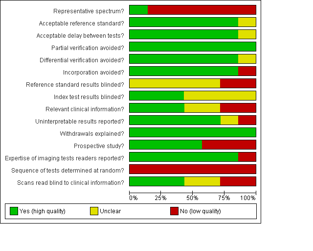
Methodological quality of the seven included studies on ischaemic stroke

Methodological quality summary: review authors' judgment on each individual QUADAS item for the seven included studies on ischaemic stroke.

Methodological quality summary: review authors' judgment on each individual QUADAS item for the two included studies on haemorrhagic stroke.

Forest plots of DWI and CT results for ischaemic stroke
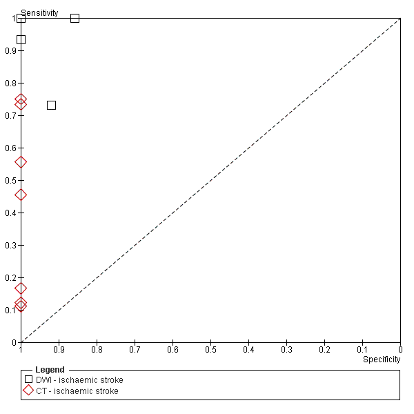
ROC plot for the seven studies that compared DWI with CT for the early detection of ischaemic stroke

MRI results for haemorrhagic stroke
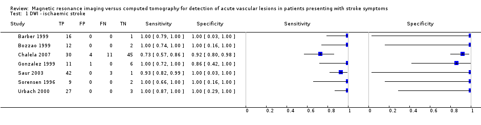
DWI ‐ ischaemic stroke.

CT ‐ ischaemic stroke.


| Review question: Comparison of diffusion‐weighted magnetic resonance imaging with conventional computer tomography for the early detection of ischaemic brain lesions in patients suspected of stroke Patient population: adults suspected of acute stroke Setting: hospital departments Geographical location: studies were conducted in Europe (3 studies), the USA (3 studies), and in Australia (1 study) Index test: diffusion‐weighted magnetic resonance imaging (DWI) performed within 12 hours of stroke onset Alternative test: computer tomography (CT) performed within 12 hours of stroke onset Reference standard: clinical assessment and imaging follow up Included studies: 7 comparative studies that evaluated DWI and CT in the same patients Total number of patients assessed: 226
| ||
| Limitations of included studies
| ||
| CT results TP 73 FP 0 FN 88 TN 65 Total 226
| DWI results TP 147 FP 5 FN 14 TN 60 Total 226 | Summary effect (95% CI) DWI sensitivity 0.99 (0.23 to 1.00) DWI specificity 0.92 (0.83 to 0.97) CT sensitivity 0.39 (0.16 to 0.69) CT specificity 1.00 (0.94 to 1.00)
|
| Conclusions and comments The small amount of data and the presence of methodological biases preclude any reliable calculation ‐ from the sensitivity and specificity estimates of CT and DWI ‐ of a positive or negative stroke diagnosis at different rates of stroke prevalence. | ||
| Applicability of tests in clinical practice | ||
| Costs | ||
| CI: confidence interval | ||
| Study | Participants (% men) | Participants assessed | Age (range) | Stroke severity | Time of imaging | MRI results (95% CI) |
| 450 (unknown) | 90 | Median 76 years (21 t0 100 years) | Median score at NIHSS = 3 (range 0 to 37) | Within 3 hours of stroke onset | GRE and DWI sensitivity 0.83 (0.52 to 0.98) GRE and DWI specificity 1.00 (0.95 to 1.00) | |
| 86 (64) | 82 | Mean 68.8 years | Mean score at NIHSS = 11.25 | Within 6 hours of stroke onset (mean time 2.6 hours) | DWI sensitivity 1.00 (0.91 to 1.00) DWI specificity 1.00 (0.91 to 1.00) GRE sensitivity 1.00 (0.91 to 1.00) GRE specificity 0.98 (0.87 to 1.00) | |
| *: prospective | ||||||
| Test | No. of studies | No. of participants |
| 1 DWI ‐ ischaemic stroke Show forest plot | 7 | 226 |
| 2 CT ‐ ischaemic stroke Show forest plot | 7 | 226 |
| 3 GRE/DWI Show forest plot | 1 | 90 |
| 4 DWI Show forest plot | 1 | 82 |
| 5 GRE Show forest plot | 1 | 82 |