Scolaris Content Display Scolaris Content Display

Risk of bias graph: review authors' judgements about each risk of bias item presented as percentages across all included studies.
Figuras y tablas -
Figure 1

Risk of bias graph: review authors' judgements about each risk of bias item presented as percentages across all included studies.

Risk of bias summary: review authors' judgements about each risk of bias item for each included study.
Figuras y tablas -
Figure 2

Risk of bias summary: review authors' judgements about each risk of bias item for each included study.

Comparison 1 Lung cancer screening with chest radiography +/‐ sputum cytology versus less intense screening, Outcome 1 Lung cancer mortality.
Figuras y tablas -
Analysis 1.1

Comparison 1 Lung cancer screening with chest radiography +/‐ sputum cytology versus less intense screening, Outcome 1 Lung cancer mortality.

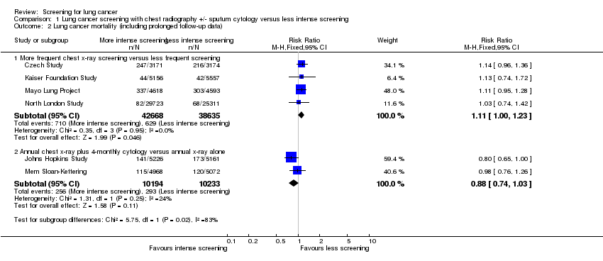
Comparison 1 Lung cancer screening with chest radiography +/‐ sputum cytology versus less intense screening, Outcome 2 Lung cancer mortality (including prolonged follow‐up data).
Figuras y tablas -
Analysis 1.2

Comparison 1 Lung cancer screening with chest radiography +/‐ sputum cytology versus less intense screening, Outcome 2 Lung cancer mortality (including prolonged follow‐up data).

Comparison 1 Lung cancer screening with chest radiography +/‐ sputum cytology versus less intense screening, Outcome 3 All‐cause mortality.
Figuras y tablas -
Analysis 1.3

Comparison 1 Lung cancer screening with chest radiography +/‐ sputum cytology versus less intense screening, Outcome 3 All‐cause mortality.

Comparison 1 Lung cancer screening with chest radiography +/‐ sputum cytology versus less intense screening, Outcome 4 Lung cancer 5‐year survival.
Figuras y tablas -
Analysis 1.4

Comparison 1 Lung cancer screening with chest radiography +/‐ sputum cytology versus less intense screening, Outcome 4 Lung cancer 5‐year survival.

Comparison 2 Annual chest x‐ray screening versus usual care (no regular screening), Outcome 1 Lung cancer mortality at 6 years of follow up.
Figuras y tablas -
Analysis 2.1

Comparison 2 Annual chest x‐ray screening versus usual care (no regular screening), Outcome 1 Lung cancer mortality at 6 years of follow up.

Comparison 2 Annual chest x‐ray screening versus usual care (no regular screening), Outcome 2 Lung cancer mortality at 13 years of follow up.
Figuras y tablas -
Analysis 2.2

Comparison 2 Annual chest x‐ray screening versus usual care (no regular screening), Outcome 2 Lung cancer mortality at 13 years of follow up.

Comparison 2 Annual chest x‐ray screening versus usual care (no regular screening), Outcome 3 Deaths from all causes (excluding deaths from PLCO cancers).
Figuras y tablas -
Analysis 2.3

Comparison 2 Annual chest x‐ray screening versus usual care (no regular screening), Outcome 3 Deaths from all causes (excluding deaths from PLCO cancers).

Comparison 3 Annual low dose CT screening versus annual chest x‐ray, Outcome 1 Lung cancer mortality.
Figuras y tablas -
Analysis 3.1

Comparison 3 Annual low dose CT screening versus annual chest x‐ray, Outcome 1 Lung cancer mortality.

Comparison 3 Annual low dose CT screening versus annual chest x‐ray, Outcome 2 All‐cause mortality.
Figuras y tablas -
Analysis 3.2

Comparison 3 Annual low dose CT screening versus annual chest x‐ray, Outcome 2 All‐cause mortality.

Summary of findings for the main comparison. Lung cancer screening with chest radiography +/‐ sputum cytology versus less intense screening for lung cancer

L ung cancer screening with chest radiography +/‐ sputum cytology versus less intense screening for lung cancer

Patient or population: Patients with lung cancer
Settings:
Intervention: Lung cancer screening with chest radiography +/‐ sputum cytology versus less intense screening

Outcomes

Illustrative comparative risks* (95% CI)

Relative effect
(95% CI)

No of Participants
(studies)

Quality of the evidence
(GRADE)

Comments

Assumed risk

Corresponding risk

Control

Lung cancer screening with chest radiography +/‐ sputum cytology versus less intense screening

Lung cancer mortality ‐ More frequent chest x‐ray screening versus less frequent screening

7 per 1000

8 per 1000
(7 to 9)

RR 1.11
(0.95 to 1.31)

81303
(4 studies)

⊕⊕⊕⊝
moderate1

Lung cancer mortality ‐ Annual chest x‐ray plus 4‐monthly cytology versus annual x‐ray alone

29 per 1000

25 per 1000
(21 to 29)

RR 0.88
(0.74 to 1.03)

20427
(2 studies)

⊕⊕⊕⊕
high

All‐cause mortality ‐ More frequent chest x‐ray screening versus less frequent screening

83 per 1000

84 per 1000
(78 to 90)

RR 1.01
(0.94 to 1.08)

170149
(4 studies)

⊕⊕⊝⊝
low2,3

All‐cause mortality ‐ Annual chest x‐ray plus 4‐monthly cytology versus annual x‐ray alone

97 per 1000

100 per 1000
(88 to 111)

RR 1.03
(0.91 to 1.15)

10040
(1 study)

⊕⊕⊕⊕
high

Lung cancer 5‐year survival ‐ More frequent chest x‐ray screening versus less frequent screening

902 per 1000

820 per 1000
(784 to 857)

RR 0.91
(0.84 to 0.99)

1775
(4 studies)

⊕⊕⊝⊝
low4,5

Lung cancer 5‐year survival ‐ Annual chest x‐ray plus 4‐monthly cytology versus annual x‐ray alone

700 per 1000

581 per 1000
(525 to 644)

RR 0.83
(0.75 to 0.92)

837
(1 study)

⊕⊕⊕⊝
moderate6

*The basis for the assumed risk (e.g. the median control group risk across studies) is provided in footnotes. The corresponding risk (and its 95% confidence interval) is based on the assumed risk in the comparison group and the relative effect of the intervention (and its 95% CI).
CI: Confidence interval; RR: Risk ratio;

GRADE Working Group grades of evidence
High quality: Further research is very unlikely to change our confidence in the estimate of effect.
Moderate quality: Further research is likely to have an important impact on our confidence in the estimate of effect and may change the estimate.
Low quality: Further research is very likely to have an important impact on our confidence in the estimate of effect and is likely to change the estimate.
Very low quality: We are very uncertain about the estimate.

1 No trials had evidence of adequate allocation concealment and only half had adequate description of drop‐outs.
2 Only half of the trials had clearly reported randomisation and there was no evidence of allocation concealment; only half of the studies had descriptions of drop‐outs.
3 I² = 56% ‐ considerable heterogeneity.
4 No evidence of allocation concealment and only one study had clear evidence of blinding.
5 I² = 68% ‐ substantial heterogeneity.
6 Single study with unclear allocation concealment and unclear risk of bias from drop‐outs.

Figuras y tablas -
Summary of findings for the main comparison. Lung cancer screening with chest radiography +/‐ sputum cytology versus less intense screening for lung cancer
Summary of findings 2. Annual chest x‐ray screening versus usual care (no regular screening) for lung cancer

Annual chest x‐ray screening versus usual care (no regular screening) for lung cancer

Patient or population: Patients with lung cancer
Settings:
Intervention: Annual chest x‐ray screening versus usual care (no regular screening)

Outcomes

Illustrative comparative risks* (95% CI)

Relative effect
(95% CI)

No of Participants
(studies)

Quality of the evidence
(GRADE)

Comments

Assumed risk

Corresponding risk

Control

Annual chest x‐ray screening versus usual care (no regular screening)

Lung cancer mortality at 6 years of follow‐up

7 per 1000

6 per 1000
(6 to 7)

RR 0.91
(0.81 to 1.03)

154901
(1 study)

⊕⊕⊕⊕
high

Lung cancer mortality at 13 years of follow‐up

16 per 1000

16 per 1000
(14 to 17)

RR 0.99
(0.91 to 1.07)

154901
(1 study)

⊕⊕⊕⊕
high

Deaths from all causes (excluding deaths from PLCO cancers)

119 per 1000

117 per 1000
(115 to 121)

RR 0.98
(0.96 to 1.01)

154901
(1 study)

⊕⊕⊕⊕
high

*The basis for the assumed risk (e.g. the median control group risk across studies) is provided in footnotes. The corresponding risk (and its 95% confidence interval) is based on the assumed risk in the comparison group and the relative effect of the intervention (and its 95% CI).
CI: Confidence interval; RR: Risk ratio;

GRADE Working Group grades of evidence
High quality: Further research is very unlikely to change our confidence in the estimate of effect.
Moderate quality: Further research is likely to have an important impact on our confidence in the estimate of effect and may change the estimate.
Low quality: Further research is very likely to have an important impact on our confidence in the estimate of effect and is likely to change the estimate.
Very low quality: We are very uncertain about the estimate.

Figuras y tablas -
Summary of findings 2. Annual chest x‐ray screening versus usual care (no regular screening) for lung cancer
Summary of findings 3. Annual low‐dose CT screening versus annual chest x‐ray for lung cancer

Annual low dose CT screening versus annual chest x‐ray for lung cancer

Patient or population: Patients with lung cancer
Settings:
Intervention: Annual low‐dose CT screening versus annual chest x‐ray

Outcomes

Illustrative comparative risks* (95% CI)

Relative effect
(95% CI)

No of Participants
(studies)

Quality of the evidence
(GRADE)

Comments

Assumed risk

Corresponding risk

Control

Annual low‐dose CT screening versus annual chest x‐ray

Lung cancer mortality

17 per 1000

13 per 1000
(12 to 15)

RR 0.8
(0.7 to 0.92)

53454
(1 study)

⊕⊕⊕⊕
high

All‐cause mortality

75 per 1000

70 per 1000
(66 to 75)

RR 0.94
(0.88 to 1)

53454
(1 study)

⊕⊕⊕⊕
high

*The basis for the assumed risk (e.g. the median control group risk across studies) is provided in footnotes. The corresponding risk (and its 95% confidence interval) is based on the assumed risk in the comparison group and the relative effect of the intervention (and its 95% CI).
CI: Confidence interval; RR: Risk ratio;

GRADE Working Group grades of evidence
High quality: Further research is very unlikely to change our confidence in the estimate of effect.
Moderate quality: Further research is likely to have an important impact on our confidence in the estimate of effect and may change the estimate.
Low quality: Further research is very likely to have an important impact on our confidence in the estimate of effect and is likely to change the estimate.
Very low quality: We are very uncertain about the estimate.

Figuras y tablas -
Summary of findings 3. Annual low‐dose CT screening versus annual chest x‐ray for lung cancer
Table 1. Number of lung cancer cases diagnosed by screening group

Study

Intervention n (%)

Intervention N

Control n(%)

Control N

Relative risk

Czech Study

108 (3.4%)

3171

82 (2.6%)

3174

1.33 (0.99,1.75)

Erfurt County Study

374 (0.9%)

41532

667 (0.7%)

102348

1.38 (1.22,1.57)

Mayo Lung Project*

585 (12.7%)

4618

500 (10.9%)

4593

1.16 (1.04,1.3)

North London Study

132 (0.44%)

29723

97 (0.38%)

25311

1.16 (0.89,1.51)

Johns Hopkins Study

238 (4.6%)

5226

246 (4.8%)

5161

0.95 (0.8,1.14)

Mem Sloan‐Kettering

176 (3.5%)

4968

178 (3.5%)

5072

1.01 (0.82,1.24)

PLCO Trial

1696 (2.2%)

77445

1620 (2.1%)

77456

1.05 (0.98, 1.12)

North American NLST

1060 (4.0%)

26722

941 (3.5%)

26732

1.13 (1.03, 1.23)

*Data from prolonged period of follow‐up reported post‐study.

Figuras y tablas -
Table 1. Number of lung cancer cases diagnosed by screening group
Comparison 1. Lung cancer screening with chest radiography +/‐ sputum cytology versus less intense screening

Outcome or subgroup title

No. of studies

No. of participants

Statistical method

Effect size

1 Lung cancer mortality Show forest plot

6

Risk Ratio (M‐H, Fixed, 95% CI)

Subtotals only

1.1 More frequent chest x‐ray screening versus less frequent screening

4

81303

Risk Ratio (M‐H, Fixed, 95% CI)

1.11 [0.95, 1.31]

1.2 Annual chest x‐ray plus 4‐monthly cytology versus annual x‐ray alone

2

20427

Risk Ratio (M‐H, Fixed, 95% CI)

0.88 [0.74, 1.03]

2 Lung cancer mortality (including prolonged follow‐up data) Show forest plot

6

Risk Ratio (M‐H, Fixed, 95% CI)

Subtotals only

2.1 More frequent chest x‐ray screening versus less frequent screening

4

81303

Risk Ratio (M‐H, Fixed, 95% CI)

1.11 [1.00, 1.23]

2.2 Annual chest x‐ray plus 4‐monthly cytology versus annual x‐ray alone

2

20427

Risk Ratio (M‐H, Fixed, 95% CI)

0.88 [0.74, 1.03]

3 All‐cause mortality Show forest plot

5

Risk Ratio (M‐H, Random, 95% CI)

Subtotals only

3.1 More frequent chest x‐ray screening versus less frequent screening

4

170149

Risk Ratio (M‐H, Random, 95% CI)

1.01 [0.94, 1.08]

3.2 Annual chest x‐ray plus 4‐monthly cytology versus annual x‐ray alone

1

10040

Risk Ratio (M‐H, Random, 95% CI)

1.03 [0.91, 1.15]

4 Lung cancer 5‐year survival Show forest plot

5

Risk Ratio (M‐H, Random, 95% CI)

Subtotals only

4.1 More frequent chest x‐ray screening versus less frequent screening

4

1775

Risk Ratio (M‐H, Random, 95% CI)

0.91 [0.84, 0.99]

4.2 Annual chest x‐ray plus 4‐monthly cytology versus annual x‐ray alone

1

837

Risk Ratio (M‐H, Random, 95% CI)

0.83 [0.75, 0.92]

Figuras y tablas -
Comparison 1. Lung cancer screening with chest radiography +/‐ sputum cytology versus less intense screening
Comparison 2. Annual chest x‐ray screening versus usual care (no regular screening)

Outcome or subgroup title

No. of studies

No. of participants

Statistical method

Effect size

1 Lung cancer mortality at 6 years of follow up Show forest plot

1

154901

Risk Ratio (M‐H, Fixed, 95% CI)

0.91 [0.81, 1.03]

2 Lung cancer mortality at 13 years of follow up Show forest plot

1

154901

Risk Ratio (M‐H, Fixed, 95% CI)

0.99 [0.91, 1.07]

3 Deaths from all causes (excluding deaths from PLCO cancers) Show forest plot

1

154901

Risk Ratio (M‐H, Fixed, 95% CI)

0.98 [0.96, 1.01]

Figuras y tablas -
Comparison 2. Annual chest x‐ray screening versus usual care (no regular screening)
Comparison 3. Annual low dose CT screening versus annual chest x‐ray

Outcome or subgroup title

No. of studies

No. of participants

Statistical method

Effect size

1 Lung cancer mortality Show forest plot

1

53454

Risk Ratio (M‐H, Fixed, 95% CI)

0.80 [0.70, 0.92]

2 All‐cause mortality Show forest plot

1

53454

Risk Ratio (M‐H, Fixed, 95% CI)

0.94 [0.88, 1.00]

Figuras y tablas -
Comparison 3. Annual low dose CT screening versus annual chest x‐ray