Contenido relacionado
Revisiones y protocolos relacionados
Rosa G Simonettia, Giovanni Perriconea, Helen L Robbinsa, Narendra R Battula, Martin O Weickert, Robert Sutton, Saboor Khan | 22 octubre 2020
Chikwendu J Ede, Dimitrinka Nikolova, Martin Brand | 3 agosto 2018
Martin Brand, Leanne Prodehl, Chikwendu J Ede | 31 octubre 2018
Peng Zhu, Sitong Dong, Ping Sun, Ajay P Belgaumkar, Yi Sun, Xiang Cheng, Qichang Zheng, Tong Li | 2 agosto 2023
Maria Corina Plaz Torres, Lawrence MJ Best, Suzanne C Freeman, Danielle Roberts, Nicola J Cooper, Alex J Sutton, Davide Roccarina, Amine Benmassaoud, Laura Iogna Prat, Norman R Williams, Mario Csenar, Dominic Fritche, Tanjia Begum, Sivapatham Arunan, Maxine Tapp, Elisabeth Jane Milne, Chavdar S Pavlov, Brian R Davidson, Emmanuel Tsochatzis, Kurinchi Selvan Gurusamy | 30 marzo 2021
Alejandro G Gonzalez-Garay, Aurora E Serralde-Zúñiga, Liliana Velasco Hidalgo, Nayelli Cointa Flores García, Ma. Isabel Aguirre-Salgado | 18 enero 2024
Danielle Roberts, Lawrence MJ Best, Suzanne C Freeman, Alex J Sutton, Nicola J Cooper, Sivapatham Arunan, Tanjia Begum, Norman R Williams, Dana Walshaw, Elisabeth Jane Milne, Maxine Tapp, Mario Csenar, Chavdar S Pavlov, Brian R Davidson, Emmanuel Tsochatzis, Kurinchi Selvan Gurusamy | 10 abril 2021
Davide Roccarina, Lawrence MJ Best, Suzanne C Freeman, Danielle Roberts, Nicola J Cooper, Alex J Sutton, Amine Benmassaoud, Maria Corina Plaz Torres, Laura Iogna Prat, Mario Csenar, Sivapatham Arunan, Tanjia Begum, Elisabeth Jane Milne, Maxine Tapp, Chavdar S Pavlov, Brian R Davidson, Emmanuel Tsochatzis, Norman R Williams, Kurinchi Selvan Gurusamy | 6 abril 2021
Amine Benmassaoud, Suzanne C Freeman, Davide Roccarina, Maria Corina Plaz Torres, Alex J Sutton, Nicola J Cooper, Laura Iogna Prat, Maxine Cowlin, Elisabeth Jane Milne, Neil Hawkins, Brian R Davidson, Chavdar S Pavlov, Douglas Thorburn, Emmanuel Tsochatzis, Kurinchi Selvan Gurusamy | 16 enero 2020
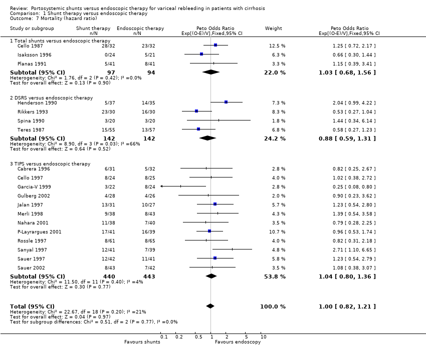
Sammy Saab, Jose M Nieto, Shannon K Lewis, Bruce A Runyon | 18 octubre 2006