Scolaris Content Display Scolaris Content Display

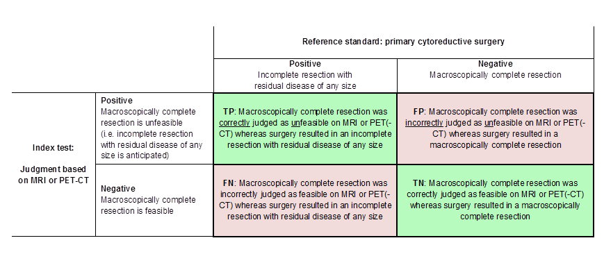
Definitions of the two by two table, wherein the index tests are tabulated against the reference standard outcome, on the analysis: macroscopic debulking versus incomplete debulking with residual disease of any size (i.e. consisting of deposits ≤ 1 cm and > 1 cm in diameter ). TP = true positive, FP = false positive, FN = false negative, TN = true negative.
Figuras y tablas -
Figure 1

Definitions of the two by two table, wherein the index tests are tabulated against the reference standard outcome, on the analysis: macroscopic debulking versus incomplete debulking with residual disease of any size (i.e. consisting of deposits ≤ 1 cm and > 1 cm in diameter ). TP = true positive, FP = false positive, FN = false negative, TN = true negative.

Definitions of the two by two table, wherein the index tests are tabulated against the reference standard outcome, on the analysis: macroscopic debulking or incomplete debulking with residual disease ≤ 1 cm in diameter versus incomplete resection with residual disease > 1 cm in diameter. TP = true positive, FP = false positive, FN = false negative, TN = true negative.
Figuras y tablas -
Figure 2

Definitions of the two by two table, wherein the index tests are tabulated against the reference standard outcome, on the analysis: macroscopic debulking or incomplete debulking with residual disease ≤ 1 cm in diameter versus incomplete resection with residual disease > 1 cm in diameter. TP = true positive, FP = false positive, FN = false negative, TN = true negative.

Visual representation of 2 x 2 table. TP = true positive, FP = false positive, FN = false negative, TN = true negative.
Figuras y tablas -
Figure 3

Visual representation of 2 x 2 table. TP = true positive, FP = false positive, FN = false negative, TN = true negative.

Study flow diagram.
Figuras y tablas -
Figure 4

Study flow diagram.

Risk of bias and applicability concerns summary: review authors' judgements about each domain for each included study
Figuras y tablas -
Figure 5

Risk of bias and applicability concerns summary: review authors' judgements about each domain for each included study

Forest plot of tests: 1 PET/CT for assessing incomplete debulking with residual disease of any size, 4 MRI for assessing incomplete debulking with residual disease of any size, 2 MRI for assessing incomplete debulking with residual disease > 1 cm, 3 MRI for assessing incomplete debulking with residual disease > 2 cm.
Figuras y tablas -
Figure 6

Forest plot of tests: 1 PET/CT for assessing incomplete debulking with residual disease of any size, 4 MRI for assessing incomplete debulking with residual disease of any size, 2 MRI for assessing incomplete debulking with residual disease > 1 cm, 3 MRI for assessing incomplete debulking with residual disease > 2 cm.

PET/CT for assessing incomplete debulking with residual disease of any size.
Figuras y tablas -
Test 1

PET/CT for assessing incomplete debulking with residual disease of any size.

MRI for assessing incomplete debulking with residual disease > 1 cm.
Figuras y tablas -
Test 2

MRI for assessing incomplete debulking with residual disease > 1 cm.

MRI for assessing incomplete debulking with residual disease > 2 cm.
Figuras y tablas -
Test 3

MRI for assessing incomplete debulking with residual disease > 2 cm.

MRI for assessing incomplete debulking with residual disease of any size.
Figuras y tablas -
Test 4

MRI for assessing incomplete debulking with residual disease of any size.

Summary of findings Diagnostic accuracy of FDG‐PET/CT and MRI for assessing tumour resectability in advanced epithelial ovarian/fallopian tube/primary peritoneal cancer

What is the diagnostic accuracy of FDG‐PET/CT or MRI for assessing tumour resectability in advanced epithelial ovarian/fallopian tube/primary peritoneal cancer?

Patients Women suspected of ovarian cancer scheduled for surgery

Prior testing Conventional diagnostic work‐up (e.g. physical examination, ultrasound)

Setting University hospitals or specialised cancer institutes

Index test FDG‐PET/CT or MRI. In all studies, the index test was evaluated as a replacement of abdominal CT. No studies were identified that followed an add‐on design.

Target condition Residual disease assessed after debulking surgery

Test

Target condition

No. of women (studies)

Prevalence in study

Sensitivity

(95% CI)

Specificity

(95% CI)

No. of false negatives*

per 1000 tested

No. of false positives**

per 1000 tested

Test accuracy certainty (quality) of evidence (sensitivity/specificity)a

FDG‐PET/CT

Residual disease > 0 cm

23/343 (2)

26%/65%

1.0 (0.54 to 1.0) and 0.66 (0.60 to 0.73)

1.0 (0.80 to 1.0) and 0.88 (0.80 to 0.93)

211 (167 to 248)b

46 (27 to 76)b

Lowc/moderated

DW‐MRI

Residual disease > 0 cm

94 (1)

53%

0.94 (0.83 to 0.99)

0.98 (0.88 to 1.00)

37 (6 to 105)b

8 (0 to 46)b

Lowc/moderated

DW‐MRI

Residual disease > 1 cm

34 (1)

23.5%

0.75 (0.35 to 0.97)

0.96 (0.80 to 1.00)

59 (7 to 153)

31 (0 to 153)

Very low/very low e, f

Conventional MRI

Residual disease > 2 cm

50 (1)

22%

0.91 (0.59 to 1.00)

0.97 (0.87 to 1.00)

20 (0 to 90)

23 (0 to 101)

Very low/very low e,g

CTh

Residual disease > 0 cm

94 (1)

53%

0.66 (95% CI 0.52 to 0.78)

0.77 (95% CI 0.63 to 0.87)

211 (136 to 298)b

87 (49 to 141)b

Low/lowc

CI: confidence interval
CT: computed tomography
DW‐MRI: diffusion‐weighted Magnetic Resonance Imaging
FDG: fluorodeoxyglucose‐18
PET: positron emission tomography
* False negatives (FNs): judged as feasible for surgery based on imaging, with an incomplete debulking at surgery.
** False positives (FPs): judged as not feasible for surgery based on imaging, with a complete debulking at surgery.

a. According to GRADE for sensitivity (false negatives (FNs)) and specificity (false positives (FPs)), respectively
b. Numbers are calculated based on the results of the largest study (Shim 2015) at the mean prevalence of incomplete debulking (62%) of the two largest studies that addressed debulking with residual disease of any size (Michielsen 2017; Shim 2015). The prevalence of incomplete debulking was calculated as (TP + FN)/total study subjects (273/437 = 62%).
c. Downgraded two levels for very wide confidence interval for number of FNs (sensitivity)
d. Downgraded one level for wide confidence interval for number of FPs (specificity)
e. Downgraded two levels as very small sample size; very wide confidence intervals for number of FNs (sensitivity) and number of FPs (specificity).
f. Downgraded one level due to applicability concerns for the Index test since the radiologists were blinded for (presurgical) clinical data.
g. Downgraded one level due to high risk of bias for patient selection and flow and timing.
h. To compare the findings of the included studies (performing PET/CT or MRI to assess tumour resectability) with CT (the current gold standard), we provided the diagnostic accuracy of CT from the study with the best quality of evidence and with the target condition that is currently used in practice (Michielsen 2017).

Figuras y tablas -
Summary of findings Diagnostic accuracy of FDG‐PET/CT and MRI for assessing tumour resectability in advanced epithelial ovarian/fallopian tube/primary peritoneal cancer
Table 1. Criteria to consider primary debulking unfeasible

Criteria to consider primary debulking unfeasible according to study methods

Alessi

Shim

Espada

Forstner

Michielsen

Site of tumour involvement

Liver/porta hepatis

Yes

No

No

Yes

Yes

Mesentery

Yes

Yes

Yes

Yes

No

Colon

Yes, when necessitating > 4 bowel resections

No

No

No

Yes, when necessitating multiple bowel resections

Stomach

Yes

No

Yes

No

Yes

Pancreas

Yes

No

No

No

Yes

Duodenum

Yes

No

No

No

Yes

Diaphragm

No

Yes

No

Yes

No

Ascites

No

Yes

No

No

No

Peritoneal carcinomatosis

Yes

Yes

No

No

No

Lesser sac/bursa omentalis

No

No

Yes

Yes

No

Spleen/splenic hilum

No

No

Yes

No

No

Lymph nodes above level of renal vessels/at coeliac axis

No

No

Yes

Yes

Yes

Gastrosplenic ligament

No

No

No

Yes

No

Presacral extraperitoneal disease

No

No

No

Yes

No

Extra‐abdominal distant metastasis

No

No

No

No

Yes

Vessels of coeliac trunk

No

No

No

No

Yes

Hepatoduodenal ligament

No

No

No

No

Yes

Superior mesenteric artery

No

No

No

No

Yes

Yes: site of tumour involvement is selected as one of the criteria to consider primary debulking unfeasible
No: site of tumour involvement is not selected as a criterion to consider primary debulking unfeasible

Figuras y tablas -
Table 1. Criteria to consider primary debulking unfeasible
Table Tests. Data tables by test

Test

No. of studies

No. of participants

1 PET/CT for assessing incomplete debulking with residual disease of any size Show forest plot

2

366

2 MRI for assessing incomplete debulking with residual disease > 1 cm Show forest plot

1

34

3 MRI for assessing incomplete debulking with residual disease > 2 cm Show forest plot

1

50

4 MRI for assessing incomplete debulking with residual disease of any size Show forest plot

1

94

Figuras y tablas -
Table Tests. Data tables by test