Scolaris Content Display Scolaris Content Display

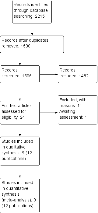
Study flow diagram.
Figuras y tablas -
Figure 1

Study flow diagram.

Risk of bias summary: review authors' judgements about each risk of bias item for each included study.
Figuras y tablas -
Figure 2

Risk of bias summary: review authors' judgements about each risk of bias item for each included study.

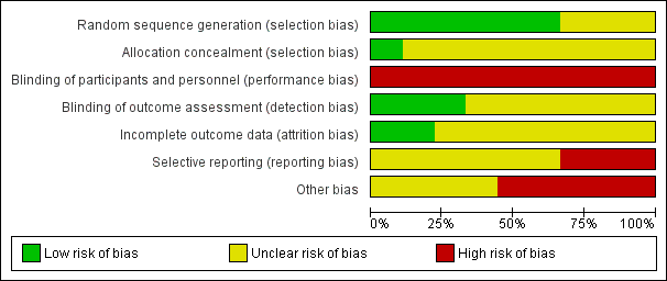
Risk of bias graph: review authors' judgements about each risk of bias item presented as percentages across all included studies.
Figuras y tablas -
Figure 3

Risk of bias graph: review authors' judgements about each risk of bias item presented as percentages across all included studies.

Forest plot of comparison: 1 Laser versus standard drill, outcome: 1.1 Caries removal (clinical).
Figuras y tablas -
Figure 4

Forest plot of comparison: 1 Laser versus standard drill, outcome: 1.1 Caries removal (clinical).

Forest plot of comparison: 1 Laser versus standard drill, outcome: 1.2 Pain.
Figuras y tablas -
Figure 5

Forest plot of comparison: 1 Laser versus standard drill, outcome: 1.2 Pain.

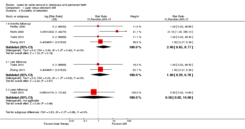
Forest plot of comparison: 1 Laser versus standard drill, outcome: 1.4 Durability of restoration.
Figuras y tablas -
Figure 6

Forest plot of comparison: 1 Laser versus standard drill, outcome: 1.4 Durability of restoration.

Forest plot of comparison: 1 Laser versus standard drill, outcome: 1.5 Pulpal inflammation or necrosis.
Figuras y tablas -
Figure 7

Forest plot of comparison: 1 Laser versus standard drill, outcome: 1.5 Pulpal inflammation or necrosis.

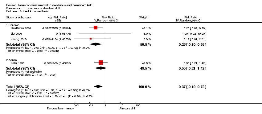
Forest plot of comparison: 1 Laser versus standard drill, outcome: 1.8 Need for anaesthesia.
Figuras y tablas -
Figure 8

Forest plot of comparison: 1 Laser versus standard drill, outcome: 1.8 Need for anaesthesia.

Comparison 1 Laser versus standard drill, Outcome 1 Caries removal (clinical).
Figuras y tablas -
Analysis 1.1

Comparison 1 Laser versus standard drill, Outcome 1 Caries removal (clinical).

Comparison 1 Laser versus standard drill, Outcome 2 Pain.
Figuras y tablas -
Analysis 1.2

Comparison 1 Laser versus standard drill, Outcome 2 Pain.

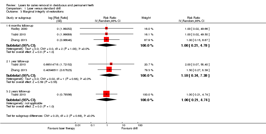
Comparison 1 Laser versus standard drill, Outcome 3 Marginal integrity of restorations.
Figuras y tablas -
Analysis 1.3

Comparison 1 Laser versus standard drill, Outcome 3 Marginal integrity of restorations.

Comparison 1 Laser versus standard drill, Outcome 4 Durability of restoration.
Figuras y tablas -
Analysis 1.4

Comparison 1 Laser versus standard drill, Outcome 4 Durability of restoration.

Comparison 1 Laser versus standard drill, Outcome 5 Pulpal inflammation or necrosis.
Figuras y tablas -
Analysis 1.5

Comparison 1 Laser versus standard drill, Outcome 5 Pulpal inflammation or necrosis.

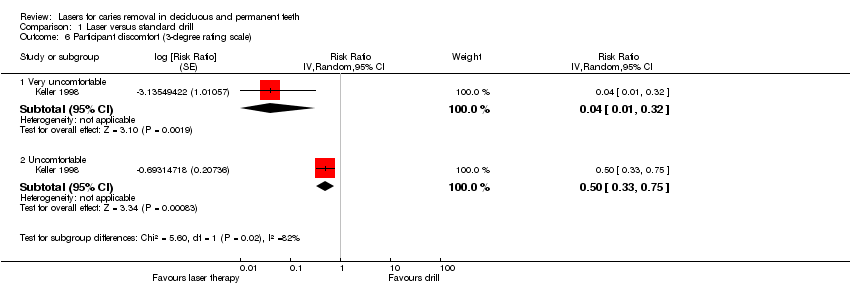
Comparison 1 Laser versus standard drill, Outcome 6 Participant discomfort (3‐degree rating scale).
Figuras y tablas -
Analysis 1.6

Comparison 1 Laser versus standard drill, Outcome 6 Participant discomfort (3‐degree rating scale).

Comparison 1 Laser versus standard drill, Outcome 7 Participant discomfort (5‐degree rating scale).
Figuras y tablas -
Analysis 1.7

Comparison 1 Laser versus standard drill, Outcome 7 Participant discomfort (5‐degree rating scale).

Comparison 1 Laser versus standard drill, Outcome 8 Need for anaesthesia.
Figuras y tablas -
Analysis 1.8

Comparison 1 Laser versus standard drill, Outcome 8 Need for anaesthesia.

Summary of findings for the main comparison. Laser compared to standard drill for caries removal in deciduous and permanent teeth

Laser compared to standard drill for caries removal in deciduous and permanent teeth

Patient or population: people with caries in deciduous and permanent teeth
Settings: primary and secondary care
Intervention: laser for caries removal
Comparison: standard drill for caries removal

Outcomes

Illustrative comparative risks* (95% CI)

Relative effect
(95% CI)

No of participants
(studies)

Quality of the evidence
(GRADE)

Comments

Assumed risk

Corresponding risk

Standard drill

Laser

Caries removal

(during treatment)

995 per 1000

995 per 1000
(990 to 1000)

RR 1.00
(0.99 to 1.01)

190 participants; 256 teeth; 256 cavity preparations
(2 studies)

⊕⊕⊝⊝
low1

Pain ‐ 6‐face rating scale (moderate and high pain)

(during treatment)

760 per 1000

304 per 1000
(236 to 395)

RR 0.40
(0.28 to 0.57)

143 participants
(2 studies)

⊕⊕⊝⊝
low2

Need for anaesthesia ‐ children

(during treatment)

97 per 1000

24 per 1000
(10 to 63)

RR 0.25
(0.10 to 0.65)

217 participants
(3 studies)

⊕⊕⊝⊝
low3

Durability of restoration ‐ 6 months follow‐up

8 per 1000

20 per 1000
(5 to 73)

RR 2.40
(0.65 to 8.77)

236 participants; 682 teeth
(4 studies)

⊕⊕⊝⊝
very low4

Marginal integrity of restorations ‐ 6 months follow‐up

7 per 1000

7 per 1000
(1 to 31)

RR 1.00
(0.21 to 4.78)

146 participants; 306 teeth
(3 studies)

⊕⊕⊝⊝
very low5

Pulpal inflammation or necrosis ‐ 1 week follow‐up

5 per 1000

7 per 1000
(1 to 36)

RR 1.51
(0.26 to 8.75)

317 participants; 694 teeth; 752 cavity preparations
(3 studies)

⊕⊕⊝⊝
very low6

Pulpal inflammation or necrosis ‐ 6 months follow‐up

4 per 1000

4 per 1000
(0 to 37)

RR 0.99
(0.10 to 9.41)

156 participants; 508 teeth; 554 cavity preparations
(2 studies)

⊕⊕⊝⊝
very low7

*The basis for the assumed risk (e.g. the median control group risk across studies) is provided in footnotes. The corresponding risk (and its 95% confidence interval) is based on the assumed risk in the comparison group and the relative effect of the intervention (and its 95% CI).
CI: confidence interval; RR: risk ratio.

GRADE Working Group grades of evidence
High quality: Further research is very unlikely to change our confidence in the estimate of effect.
Moderate quality: Further research is likely to have an important impact on our confidence in the estimate of effect and may change the estimate.
Low quality: Further research is very likely to have an important impact on our confidence in the estimate of effect and is likely to change the estimate.
Very low quality: We are very uncertain about the estimate.

1The evidence was downgraded by two levels because of very serious concern regarding the risk of bias: (1) the two studies did not report sufficient information regarding the allocation concealment (DenBesten 2001; Hadley 2000); (2) both studies were at high risk of performance bias; (3) neither of the two studies was at low risk of selective reporting bias; (4) serious concern related to funding (other bias).
2The evidence was downgraded by two levels because of very serious concern regarding the risk of bias: one study (Belcheva 2014), in addition to being at high risk of performance bias, had no item with low risk of bias; the second study was at high risk of performance bias and at unclear risk of selection bias (the method of allocation concealment was not reported) and the remaining items of the risk of bias (Zhang 2013).
3The evidence was downgraded by two levels because of very serious concern regarding the risk of bias: (1) unclear risk of selection bias (all three studies); (2) high risk of performance bias (all three studies); unclear risk of selective reporting bias (all three studies); unclear risk of attrition bias (DenBesten 2001; Zhang 2013); (3) two studies with high risk of other bias (DenBesten 2001; Liu 2006) and one with unclear risk of other bias (Zhang 2013).
4The overall evidence was downgraded by three levels: two levels because of very serious concern regarding the risk of bias (no study with low risk of bias; all studies with high risk of performance bias; three studies at unclear risk of attrition bias (Hadley 2000; Harris 2000; Zhang 2013); two studies at high risk of selective reporting bias (Harris 2000; Yazici 2010); two with unclear selective reporting bias (Hadley 2000; Zhang 2013); two with high risk of other bias (Hadley 2000; Harris 2000)); one level because of lack of precision.
5The overall evidence was downgraded by three levels: two levels because of very serious concern regarding the risk of bias (no study with low risk of bias; all studies with high risk of performance bias; one study at high risk of selective reporting bias (Yazici 2010), two with unclear selective reporting bias (Hadley 2000; Zhang 2013); one with high risk of other bias (Hadley 2000)); one level because of lack of precision.
6The overall evidence was downgraded by three levels: two levels because of very serious concern regarding the risk of bias (no study with low risk of selection bias; all studies with high risk of performance bias; one study at high risk of selective reporting bias (Harris 2000) and two at unclear risk of selective reporting bias (DenBesten 2001; Keller 1998); two studies at high risk of other bias (DenBesten 2001; Harris 2000)); one level because of lack of precision.
7The overall evidence was downgraded by three levels: two levels because of very serious concern regarding the risk of bias (no study with low risk of bias; all studies with high risk of performance bias; no study at low risk of selective reporting bias or other bias (Hadley 2000; Harris 2000)); one level because of lack of precision.

Figuras y tablas -
Summary of findings for the main comparison. Laser compared to standard drill for caries removal in deciduous and permanent teeth
Comparison 1. Laser versus standard drill

Outcome or subgroup title

No. of studies

No. of participants

Statistical method

Effect size

1 Caries removal (clinical) Show forest plot

2

Risk Ratio (Random, 95% CI)

1.00 [0.99, 1.01]

2 Pain Show forest plot

3

Risk Ratio (Random, 95% CI)

Subtotals only

2.1 6‐face rating scale (moderate and high pain)

2

Risk Ratio (Random, 95% CI)

0.40 [0.28, 0.57]

2.2 Modified simple 4‐face scale

1

Risk Ratio (Random, 95% CI)

0.21 [0.11, 0.42]

3 Marginal integrity of restorations Show forest plot

3

Risk Ratio (Random, 95% CI)

Subtotals only

3.1 6 months follow‐up

3

Risk Ratio (Random, 95% CI)

1.0 [0.21, 4.78]

3.2 1 year follow‐up

2

Risk Ratio (Random, 95% CI)

1.59 [0.34, 7.38]

3.3 2 years follow‐up

1

Risk Ratio (Random, 95% CI)

1.0 [0.21, 4.74]

4 Durability of restoration Show forest plot

4

Risk Ratio (Random, 95% CI)

Subtotals only

4.1 6 months follow‐up

4

Risk Ratio (Random, 95% CI)

2.40 [0.65, 8.77]

4.2 1 year follow‐up

2

Risk Ratio (Random, 95% CI)

1.40 [0.29, 6.78]

4.3 2 years follow‐up

1

Risk Ratio (Random, 95% CI)

0.50 [0.02, 14.60]

5 Pulpal inflammation or necrosis Show forest plot

4

Risk Ratio (Random, 95% CI)

1.29 [0.32, 5.14]

5.1 1 week

3

Risk Ratio (Random, 95% CI)

1.51 [0.26, 8.75]

5.2 6 months

2

Risk Ratio (Random, 95% CI)

0.99 [0.10, 9.41]

6 Participant discomfort (3‐degree rating scale) Show forest plot

1

Risk Ratio (Random, 95% CI)

Subtotals only

6.1 Very uncomfortable

1

Risk Ratio (Random, 95% CI)

0.04 [0.01, 0.32]

6.2 Uncomfortable

1

Risk Ratio (Random, 95% CI)

0.50 [0.33, 0.75]

7 Participant discomfort (5‐degree rating scale) Show forest plot

1

Risk Ratio (Random, 95% CI)

Subtotals only

7.1 Mild discomfort

1

Risk Ratio (Random, 95% CI)

0.12 [0.01, 2.32]

7.2 Moderate discomfort

1

Risk Ratio (Random, 95% CI)

0.33 [0.04, 3.12]

8 Need for anaesthesia Show forest plot

4

Risk Ratio (Random, 95% CI)

0.37 [0.19, 0.72]

8.1 Children

3

Risk Ratio (Random, 95% CI)

0.25 [0.10, 0.65]

8.2 Adults

1

Risk Ratio (Random, 95% CI)

0.55 [0.21, 1.42]

Figuras y tablas -
Comparison 1. Laser versus standard drill