Adhésifs utilisés pour les tubes molaires collés pendant un traitement avec un appareil fixe
Referencias
References to studies included in this review
Additional references
References to other published versions of this review
Characteristics of studies
Characteristics of included studies [ordered by study ID]
| Methods | RCT, parallel group. Multicentre: 2 UK hospital orthodontic clinics. Follow‐up: end or discontinuation of treatment. | |
| Participants | 110 hospital waiting list patients needing (with no previous history of) fixed appliances. Age: 9 to 33 years. Duration of treatment: 7 months to 41 months. | |
| Interventions | Group 1. Single first molar tubes bonded with a no‐mix chemically cured composite (Rely‐A‐Bond) after a 30 second etch (55 participants; 181 molar tubes). Group 2. Non‐sandblasted bands cemented with conventional glass ionomer cement (Intact) (55 participants; 186 bands). All participants received similar straight wire mechanics and archwire sequences. | |
| Outcomes | First time failure (detachment or loosening of the attachment). The primary outcome was attachment failure (tooth level) and secondary outcome the number of failures per patient (participant level). Time to failure. Adhesive Remnant Index. | |
| Notes | ||
| Risk of bias | ||
| Bias | Authors' judgement | Support for judgement |
| Random sequence generation (selection bias) | Low risk | Quote: "random number tables". |
| Allocation concealment (selection bias) | Low risk | Quote: "Each operator enrolled participants and assigned them to their group using their sealed [opaque] envelopes, which blinded the operator and participant to the assignment before enrolment". |
| Incomplete outcome data (attrition bias) | Low risk | Comment: all participants accounted for. |
| Selective reporting (reporting bias) | Low risk | Comment: all relevant outcomes reported. |
| Other bias | Low risk | Comment: no other bias evident. |
| Methods | RCT, parallel group. Multicentre: 3 UK orthodontic clinics. Follow‐up: end of treatment. | |
| Participants | 80 patients starting upper and lower fixed appliance (pre‐adjusted edgewise) treatment. Age: 10 to 18 years. Duration of treatment: unclear. | |
| Interventions | Group 1. Tubes bonded with light‐cured composite (3M Unitek Transbond XT) to all 4 first permanent molar teeth for each participant (38 participants analysed; 152 tubes). Group 2. Bands cemented with glass ionomer (3M ESPE Ketac‐Cem) to all 4 first permanent molar teeth for each participant (38 participants analysed; 152 bands). | |
| Outcomes | First time failure. Decalcification. | |
| Notes | ||
| Risk of bias | ||
| Bias | Authors' judgement | Support for judgement |
| Random sequence generation (selection bias) | Low risk | Quote: "random number tables". |
| Allocation concealment (selection bias) | Low risk | Quote: "allocations were concealed in envelopes marked with each subject's identification number and held in a central place. The operator and patient remained blind to the attachment type until after the consent and registration procedures". |
| Incomplete outcome data (attrition bias) | Low risk | Comment: all participants accounted for (80 randomised; 76 analysed for failure; 74 analysed for decalcification). |
| Selective reporting (reporting bias) | Low risk | Comment: all relevant outcomes reported. |
| Other bias | Low risk | Comment: no other bias evident. |
RCT = randomised controlled trial.
Data and analyses
| Outcome or subgroup title | No. of studies | No. of participants | Statistical method | Effect size |
| 1 Failure at tooth level Show forest plot | 2 | Hazard Ratio (Fixed, 95% CI) | 2.92 [1.80, 4.72] | |
| Analysis 1.1  Comparison 1 Molar tubes versus molar bands, Outcome 1 Failure at tooth level. | ||||
| 2 Failure at participant level Show forest plot | 2 | 186 | Risk Ratio (M‐H, Fixed, 95% CI) | 2.30 [1.56, 3.41] |
| Analysis 1.2  Comparison 1 Molar tubes versus molar bands, Outcome 2 Failure at participant level. | ||||
| 3 Decalcification (participant level) Show forest plot | 1 | 74 | Risk Ratio (M‐H, Random, 95% CI) | 1.85 [1.22, 2.79] |
| Analysis 1.3  Comparison 1 Molar tubes versus molar bands, Outcome 3 Decalcification (participant level). | ||||
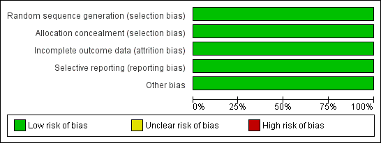
Risk of bias graph: review authors' judgements about each risk of bias item presented as percentages across all included studies.
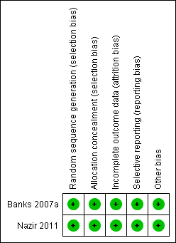
Risk of bias summary: review authors' judgements about each risk of bias item for each included study.
Comparison 1 Molar tubes versus molar bands, Outcome 1 Failure at tooth level.

Comparison 1 Molar tubes versus molar bands, Outcome 2 Failure at participant level.

Comparison 1 Molar tubes versus molar bands, Outcome 3 Decalcification (participant level).
| Outcome or subgroup title | No. of studies | No. of participants | Statistical method | Effect size |
| 1 Failure at tooth level Show forest plot | 2 | Hazard Ratio (Fixed, 95% CI) | 2.92 [1.80, 4.72] | |
| 2 Failure at participant level Show forest plot | 2 | 186 | Risk Ratio (M‐H, Fixed, 95% CI) | 2.30 [1.56, 3.41] |
| 3 Decalcification (participant level) Show forest plot | 1 | 74 | Risk Ratio (M‐H, Random, 95% CI) | 1.85 [1.22, 2.79] |