Scolaris Content Display Scolaris Content Display

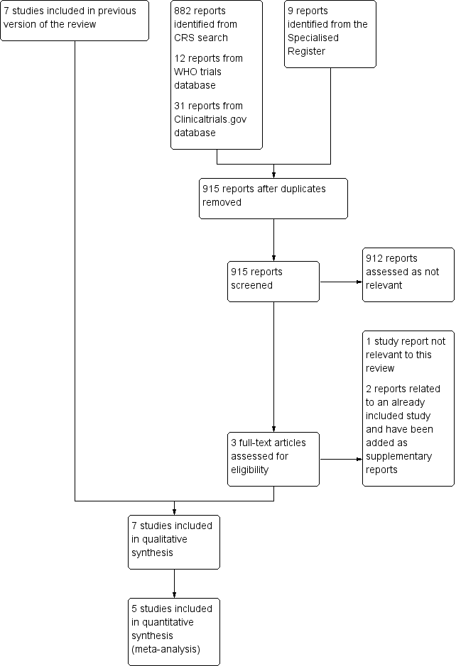
Study flow diagram.
Figures and Tables -
Figure 1

Study flow diagram.

Risk of bias graph: review authors' judgements about each risk of bias item presented as percentages across all included studies.
Figures and Tables -
Figure 2

Risk of bias graph: review authors' judgements about each risk of bias item presented as percentages across all included studies.

Risk of bias summary: review authors' judgements about each risk of bias item for each included study.
Figures and Tables -
Figure 3

Risk of bias summary: review authors' judgements about each risk of bias item for each included study.

Forest plot of comparison: 1 Clinically‐indicated versus routine change, outcome: 1.1 Catheter‐related bloodstream infection.
Figures and Tables -
Figure 4

Forest plot of comparison: 1 Clinically‐indicated versus routine change, outcome: 1.1 Catheter‐related bloodstream infection.

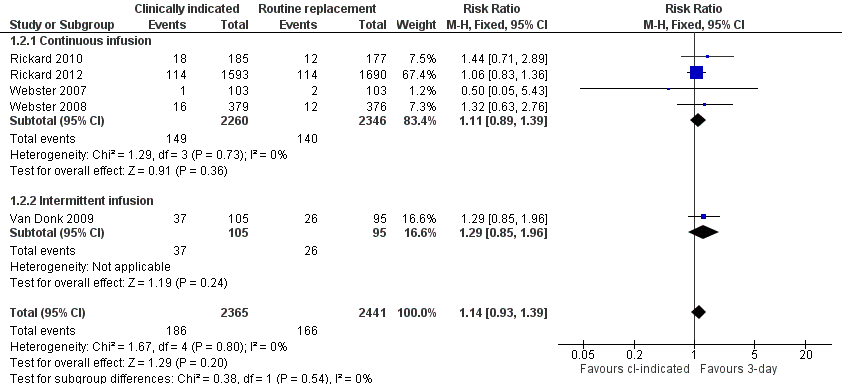
Forest plot of comparison: 1 Clinically‐indicated versus routine change, outcome: 1.2 Phlebitis.
Figures and Tables -
Figure 5

Forest plot of comparison: 1 Clinically‐indicated versus routine change, outcome: 1.2 Phlebitis.

Forest plot of comparison: 1 Clinically‐indicated versus routine change, outcome: 1.3 Phlebitis per device days.
Figures and Tables -
Figure 6

Forest plot of comparison: 1 Clinically‐indicated versus routine change, outcome: 1.3 Phlebitis per device days.

Forest plot of comparison: 1 Clinically‐indicated versus routine change, outcome: 1.4 All‐cause bloodstream infection.
Figures and Tables -
Figure 7

Forest plot of comparison: 1 Clinically‐indicated versus routine change, outcome: 1.4 All‐cause bloodstream infection.

Forest plot of comparison: 1 Clinically‐indicated versus routine change, outcome: 1.5 Cost.
Figures and Tables -
Figure 8

Forest plot of comparison: 1 Clinically‐indicated versus routine change, outcome: 1.5 Cost.

Forest plot of comparison: 1 Clinically‐indicated versus routine change, outcome: 1.6 Infiltration.
Figures and Tables -
Figure 9

Forest plot of comparison: 1 Clinically‐indicated versus routine change, outcome: 1.6 Infiltration.

Forest plot of comparison: 1 Clinically‐indicated versus routine change, outcome: 1.7 Catheter blockage.
Figures and Tables -
Figure 10

Forest plot of comparison: 1 Clinically‐indicated versus routine change, outcome: 1.7 Catheter blockage.

Forest plot of comparison: 1 Clinically‐indicated versus routine change, outcome: 1.8 Local infection.
Figures and Tables -
Figure 11

Forest plot of comparison: 1 Clinically‐indicated versus routine change, outcome: 1.8 Local infection.

Forest plot of comparison: 1 Clinically‐indicated versus routine change, outcome: 1.9 Mortality.
Figures and Tables -
Figure 12

Forest plot of comparison: 1 Clinically‐indicated versus routine change, outcome: 1.9 Mortality.

Comparison 1 Clinically‐indicated versus routine change, Outcome 1 Catheter‐related blood stream infection.
Figures and Tables -
Analysis 1.1

Comparison 1 Clinically‐indicated versus routine change, Outcome 1 Catheter‐related blood stream infection.

Comparison 1 Clinically‐indicated versus routine change, Outcome 2 Phlebitis.
Figures and Tables -
Analysis 1.2

Comparison 1 Clinically‐indicated versus routine change, Outcome 2 Phlebitis.

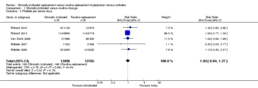
Comparison 1 Clinically‐indicated versus routine change, Outcome 3 Phlebitis per device days.
Figures and Tables -
Analysis 1.3

Comparison 1 Clinically‐indicated versus routine change, Outcome 3 Phlebitis per device days.

Comparison 1 Clinically‐indicated versus routine change, Outcome 4 All‐cause blood stream infection.
Figures and Tables -
Analysis 1.4

Comparison 1 Clinically‐indicated versus routine change, Outcome 4 All‐cause blood stream infection.

Comparison 1 Clinically‐indicated versus routine change, Outcome 5 Cost.
Figures and Tables -
Analysis 1.5

Comparison 1 Clinically‐indicated versus routine change, Outcome 5 Cost.

Comparison 1 Clinically‐indicated versus routine change, Outcome 6 Infiltration.
Figures and Tables -
Analysis 1.6

Comparison 1 Clinically‐indicated versus routine change, Outcome 6 Infiltration.

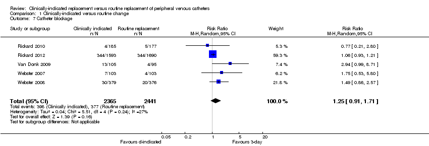
Comparison 1 Clinically‐indicated versus routine change, Outcome 7 Catheter blockage.
Figures and Tables -
Analysis 1.7

Comparison 1 Clinically‐indicated versus routine change, Outcome 7 Catheter blockage.

Comparison 1 Clinically‐indicated versus routine change, Outcome 8 Local infection.
Figures and Tables -
Analysis 1.8

Comparison 1 Clinically‐indicated versus routine change, Outcome 8 Local infection.

Comparison 1 Clinically‐indicated versus routine change, Outcome 9 Mortality.
Figures and Tables -
Analysis 1.9

Comparison 1 Clinically‐indicated versus routine change, Outcome 9 Mortality.

Summary of findings for the main comparison. Clinically‐indicated versus routine changes for peripheral venous catheter‐related complications

Clinically‐indicated versus routine changes for peripheral venous catheter‐related complications

Patient or population: patients with peripheral venous catheter‐related complications
Settings: Hospitals and community settings
Intervention: clinically‐indicated versus routine changes

Outcomes

Illustrative comparative risks* (95% CI)

Relative effect
(95% CI)

No of Participants
(studies)

Quality of the evidence
(GRADE)

Comments

Assumed risk

Corresponding risk

Control

Clinically indicated versus routine changes

Catheter‐related bloodstream infection
Positive blood culture from a peripheral vein; clinical signs of infection; no other apparent source for the bloodstream infection except the intravenous catheter; and colonised intravenous catheter tip culture with the same organism as identified in the blood

Study population

RR 0.61
(0.08 to 4.68)

4806
(5 studies)

⊕⊕⊕⊖
moderate1,2,3,4

1 per 1000

1 per 1000
(0 to 5)

Moderate

0 per 1000

0 per 1000
(0 to 0)

Phlebitis
Any definition used by the author

Study population

RR 1.14
(0.93 to 1.39)

4806
(5 studies)

⊕⊕⊕⊕
high1,3

68 per 1000

78 per 1000
(63 to 95)

Moderate

68 per 1000

78 per 1000
(63 to 95)

All‐cause bloodstream infection

Study population

RR 0.47
(0.15 to 1.53)

3283
(1 study)

⊕⊕⊕⊕
high1,3

5 per 1000

3 per 1000
(1 to 8)

Moderate

5 per 1000

2 per 1000
(1 to 8)

Cost
Estmated. Based on materials and staff costs5,6

The mean cost in the intervention groups was
AUD $6.96 lower
(9.05 to 4.86 lower)

4244
(3 studies)

⊕⊕⊕⊕
high

*The basis for the assumed risk (e.g. the median control group risk across studies) is provided in footnotes. The corresponding risk (and its 95% confidence interval) is based on the assumed risk in the comparison group and the relative effect of the intervention (and its 95% CI).
CI: Confidence interval; RR: Risk ratio;

GRADE Working Group grades of evidence
High quality: Further research is very unlikely to change our confidence in the estimate of effect.
Moderate quality: Further research is likely to have an important impact on our confidence in the estimate of effect and may change the estimate.
Low quality: Further research is very likely to have an important impact on our confidence in the estimate of effect and is likely to change the estimate.
Very low quality: We are very uncertain about the estimate.

1 Although patients and those recording outcomes were aware of group allocation, it seems unlikely that this knowledge would have affected results. None of those recording outcomes were investigators and the diagnosis was based on verifiable data in patients medical records.
2 In three of the five trials, no CRBSI occurred in either arm of the study. In the other two trials there was considerable overlap in the confidence intervals, consequently there was no statistical heterogeneity.
3 Participants, interventions and outcomes were similar across studies.
4 Confidence intervals were wide for this outcome, indicating a level of uncertainty around the effect size.
5 The overall cost for cannula replacement varies by cost of materials, time, solutions, additives to the solution.
6 Mean cost is reported in Australian dollars.

Figures and Tables -
Summary of findings for the main comparison. Clinically‐indicated versus routine changes for peripheral venous catheter‐related complications
Comparison 1. Clinically‐indicated versus routine change

Outcome or subgroup title

No. of studies

No. of participants

Statistical method

Effect size

1 Catheter‐related blood stream infection Show forest plot

5

4806

Risk Ratio (M‐H, Fixed, 95% CI)

0.61 [0.08, 4.68]

2 Phlebitis Show forest plot

5

4806

Risk Ratio (M‐H, Fixed, 95% CI)

1.14 [0.93, 1.39]

2.1 Continuous infusion

4

4606

Risk Ratio (M‐H, Fixed, 95% CI)

1.11 [0.89, 1.39]

2.2 Intermittent infusion

1

200

Risk Ratio (M‐H, Fixed, 95% CI)

1.29 [0.85, 1.96]

3 Phlebitis per device days Show forest plot

5

26191

Risk Ratio (M‐H, Fixed, 95% CI)

1.03 [0.84, 1.27]

4 All‐cause blood stream infection Show forest plot

1

Risk Ratio (M‐H, Fixed, 95% CI)

Totals not selected

5 Cost Show forest plot

3

4244

Mean Difference (IV, Fixed, 95% CI)

‐6.96 [‐9.05, ‐4.86]

6 Infiltration Show forest plot

4

4606

Risk Ratio (M‐H, Fixed, 95% CI)

1.17 [1.05, 1.31]

7 Catheter blockage Show forest plot

5

4806

Risk Ratio (M‐H, Random, 95% CI)

1.25 [0.91, 1.71]

8 Local infection Show forest plot

4

4606

Risk Ratio (M‐H, Fixed, 95% CI)

4.96 [0.24, 102.98]

9 Mortality Show forest plot

1

Risk Ratio (M‐H, Fixed, 95% CI)

Totals not selected

Figures and Tables -
Comparison 1. Clinically‐indicated versus routine change